. 2015 May 8:6:7062.
doi: 10.1038/ncomms8062.

Ventral hippocampal afferents to the nucleus accumbens regulate susceptibility to depression

Affiliations

Ventral hippocampal afferents to the nucleus accumbens regulate susceptibility to depression

Rosemary C Bagot et al. Nat Commun. .

Erratum in

Abstract

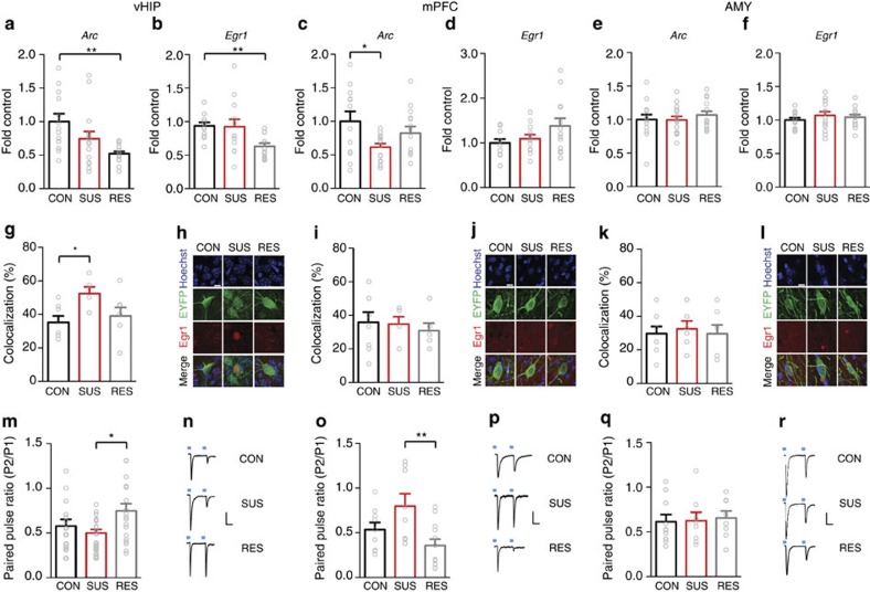
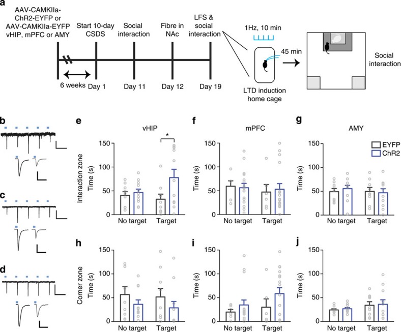
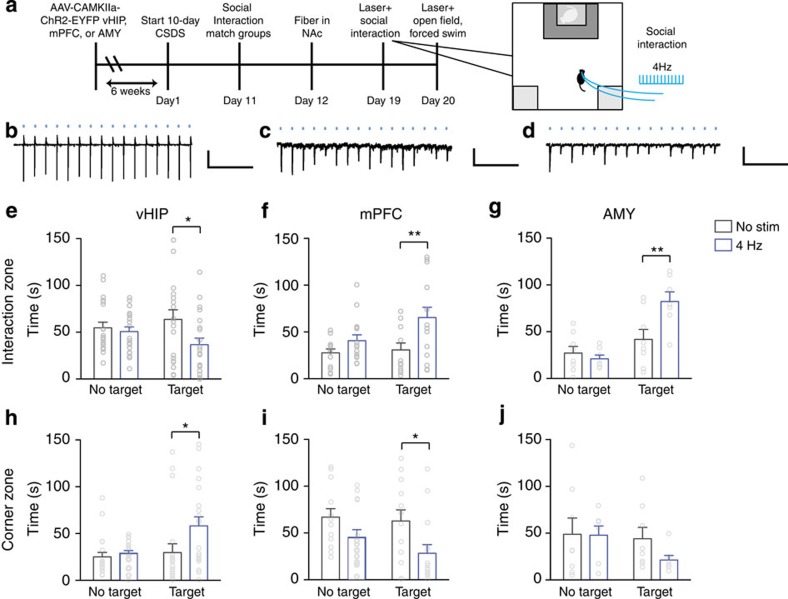
Enhanced glutamatergic transmission in the nucleus accumbens (NAc), a region critical for reward and motivation, has been implicated in the pathophysiology of depression; however, the afferent source of this increased glutamate tone is not known. The NAc receives glutamatergic inputs from the medial prefrontal cortex (mPFC), ventral hippocampus (vHIP) and basolateral amygdala (AMY). Here, we demonstrate that glutamatergic vHIP afferents to NAc regulate susceptibility to chronic social defeat stress (CSDS). We observe reduced activity in vHIP in mice resilient to CSDS. Furthermore, attenuation of vHIP-NAc transmission by optogenetic induction of long-term depression is pro-resilient, whereas acute enhancement of this input is pro-susceptible. This effect is specific to vHIP afferents to the NAc, as optogenetic stimulation of either mPFC or AMY afferents to the NAc is pro-resilient. These data indicate that vHIP afferents to NAc uniquely regulate susceptibility to CSDS, highlighting an important, novel circuit-specific mechanism in depression.


Figures

Figure 1
Figure 1. Opposing effects of CSDS on vHIP and mPFC IEG expression and synaptic function.
mRNA expression of two immediate early genes in vHIP of control (CON), susceptible (SUS) and resilient (RES) mice. (a) Arc (F2,40=6.485, P<0.01, post hoc **P<0.01, n=13,15,15) and (b) Egr1 (F2,36=5.415, P<0.01, post hoc **P<0.01, n=12,14,13) were decreased in vHIP of RES mice 48 h post-defeat. In contrast, (c) Arc expression was decreased in mPFC of SUS mice (F2,37=3.431, P<0.05, post hoc CON versus SUS *P<0.05, n=12,14,14). (d) Egr1 expression was not significantly different (n=11,13,12). Neither Arc (e) nor Egr1 (f) expression was regulated by defeat in AMY (n=14,15,15). A greater percentage of EYFP retrogradely labelled NAc-projecting neurons were EGR1 positive in vHIP (g) of SUS mice compared to controls (F2,16=3.618, P<0.05, post hoc *P<0.05, n=7,5,7). EGR1 levels in mPFC (i) and AMY (k) in NAc-projecting neurons were not significantly regulated by defeat. Representative images show Hoecsht nuclear stain (blue) in EYFP labelled EGR1 (red) staining in NAc-projecting neurons in vHIP (h), mPFC (j) and AMY (l) labelled by retrograde AAV2/5–CaMKIIa–EYFP injected into NAc (scale bar 10 μm). In RES relative to SUS mice, paired-pulse ratios (P2/P1; 100 ms interpulse interval) of optically evoked EPSCs in NAc shell MSNs were increased from (m) vHIP (F2,50=4.023, P<0.05, post hoc *P<0.05, n=14,20,19 cells from n=6,7,6 mice) and decreased from (o) mPFC (F2,26=5.459, P<0.01, post hoc **P<0.01, n=9,8,12 cells from n=5,4,7 mice). Paired-pulse ratios from AMY (q) were not regulated by defeat (n=10,8,8 cells from n=4,3,4 mice). Representative EPSCs evoked by optical stimulation of vHIP (n), mPFC (p) and AMY (r) ChR2-expressing terminals. Vertical scale bar (n) 80 pA, (p,r) 40 pA, horizontal scale bar 40 ms. Blue squares indicate light pulse delivery. One-way-ANOVAs with Bonferroni post hoc tests. Grubb's test was used to detect and remove statistical outliers in panels a-f. Error bars represent s.e.m.
Figure 2
Figure 2. LTD at vHIP-NAc induces resilience to CSDS.
(a). Schematic of experimental time-course. Representative traces validating that 1 Hz stimulation of ChR2-expressing vHIP (b) (scale bar 200 pA, 1 s), mPFC (c) (scale bar 50 pA, 1 s) and AMY (d) (scale bar 200 pA, 1 s) terminals reliably evoke EPSCs with temporal fidelity in NAc MSNs (upper panels). 10 min, 1 Hz stimulation (low frequency stimulation; LFS) induces LTD of evoked EPSCs measured 45 min after LFS (bd, lower panels; scale bars 80 pA, 50 ms) 45 min post-LFS (grey traces) compared to pre-LFS baselines (black traces). Blue squares indicate light pulse delivery. (e) In vivo, LFS of vHIP-NAc synapses in defeated mice increased time spent in the interaction zone in the presence of a target (F1,18=5.274 interaction effect, P<0.05, post hoc *P<0.05, n=9,11) but did not alter time spent in corners (h). Stimulation of mPFC-NAc (f; n=6, 16) or AMY-NAc synapses (g; n=9,11) had no effect on time spent in interaction or corner zones (i, j). Two-way repeated measures ANOVA with Bonferroni post hoc tests. ‘EYFP' and ‘ChR2' denote AAV5-CaMKIIa–EYFP and AAV5-CaMKIIa–ChR2–EYFP, respectively. ‘No target' and ‘Target' indicate absence or presence of a target mouse during testing. Error bars represent s.e.m.
Figure 3
Figure 3. Acute stimulation of vHIP versus mPFC or AMY terminals in NAc promotes opposite behavioral responses to CSDS.
(a). Schematic of experimental time course. Representative traces validating that 4 Hz stimulation of vHIP (b) (scale bar 200 pA, 1 s), mPFC (c) (scale bar 50 pA, 1 s) and AMY (d) (scale bar 50 pA, 1 s) terminals in NAc slices evokes EPSCs. Blue squares indicate light pulse delivery. In vivo, 4 Hz stimulation of vHIP terminals in NAc shell (e) reduced time spent interacting with a target mouse (F1,39=4.274 interaction effect, P<0.05, post hoc *P<0.05, n=20,21) and (h) increased time spent in corners (F1,39=6.560 target effect, F1,39=4.271, stimulation effect, P<0.05, post hoc *P<0.05, n=20,21). Stimulation of mPFC terminals in NAc (f) increased time spent interacting with a target mouse (F1,26=9.174 stimulation effect, P<0.01, post hoc **P<0.01, n=13,15) and (i) decreased time spent in corners (F1,26=7.735, P<0.01, post hoc *P<0.05, n=13,15). Stimulation of AMY axons in NAc (g) increased time spent interacting with a target mouse (F1,14=5.4284 interaction effect, P<0.01, post hoc **P<0.01, n=8) but (j) did not affect time spent in corners. Two-way repeated-measures ANOVAs with Bonferroni post hoc tests. ‘No stim' and ‘4 Hz' denote non-stimulated controls versus 4 Hz stimulated mice. ‘No target' and ‘Target' indicate absence or presence of a target mouse during testing. Error bars represent s.e.m.

References

    1. Krishnan V. et al.. Molecular adaptations underlying susceptibility and resistance to social defeat in brain reward regions. Cell 131, 391–404 (2007). - PubMed
    1. Berton O. et al.. Essential role of BDNF in the mesolimbic dopamine pathway in social defeat stress. Science 311, 864–868 (2006). - PubMed
    1. Vialou V. et al.. DeltaFosB in brain reward circuits mediates resilience to stress and antidepressant responses. Nat. Neurosci. 13, 745–752 (2010). - PMC - PubMed
    1. Christoffel D. J. et al.. IkappaB kinase regulates social defeat stress-induced synaptic and behavioral plasticity. J. Neurosci. 31, 314–321 (2011). - PMC - PubMed
    1. Lim B. K., Huang K. W., Grueter B. A., Rothwell P. E. & Malenka R. C. Anhedonia requires MC4R-mediated synaptic adaptations in nucleus accumbens. Nature 487, 183–189 (2012). - PMC - PubMed

Publication types