. 2015 May 8:13:152.
doi: 10.1186/s12967-015-0499-8.

Osteoinduction and proliferation of bone-marrow stromal cells in three-dimensional poly (ε-caprolactone)/ hydroxyapatite/collagen scaffolds

Affiliations

Osteoinduction and proliferation of bone-marrow stromal cells in three-dimensional poly (ε-caprolactone)/ hydroxyapatite/collagen scaffolds

Ting Wang et al. J Transl Med. .

Abstract

Background: Osteoinduction and proliferation of bone-marrow stromal cells (BMSCs) in three-dimensional (3D) poly(ε-caprolactone) (PCL) scaffolds have not been studied throughly and are technically challenging. This study aimed to optimize nanocomposites of 3D PCL scaffolds to provide superior adhesion, proliferation and differentiation environment for BMSCs in this scenario.

Methods: BMSCs were isolated and cultured in a novel 3D tissue culture poly(ε-caprolactone) (PCL) scaffold coated with poly-lysine, hydroxyapatite (HAp), collagen and HAp/collagen. Cell morphology was observed and BMSC biomarkers for osteogenesis, osteoblast differentiation and activation were analyzed.

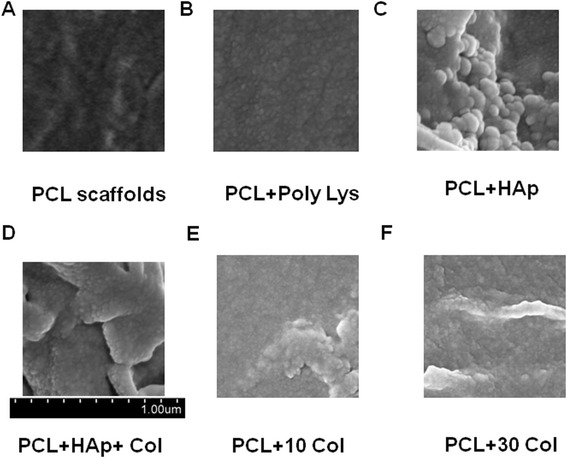
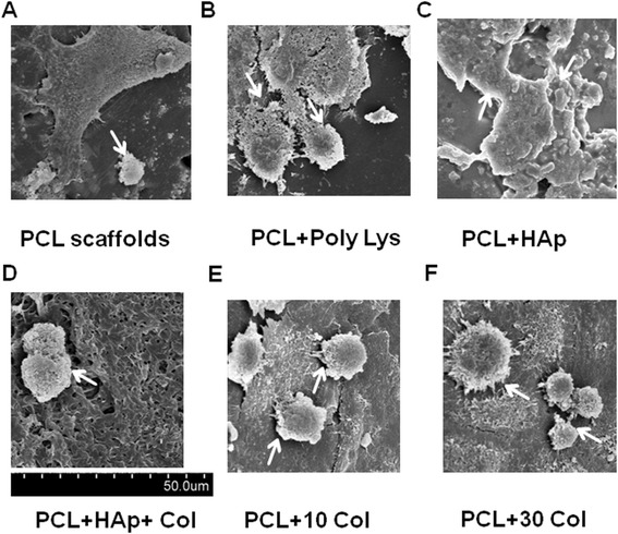
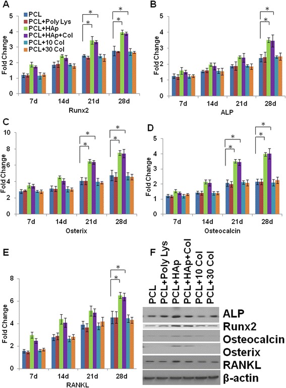
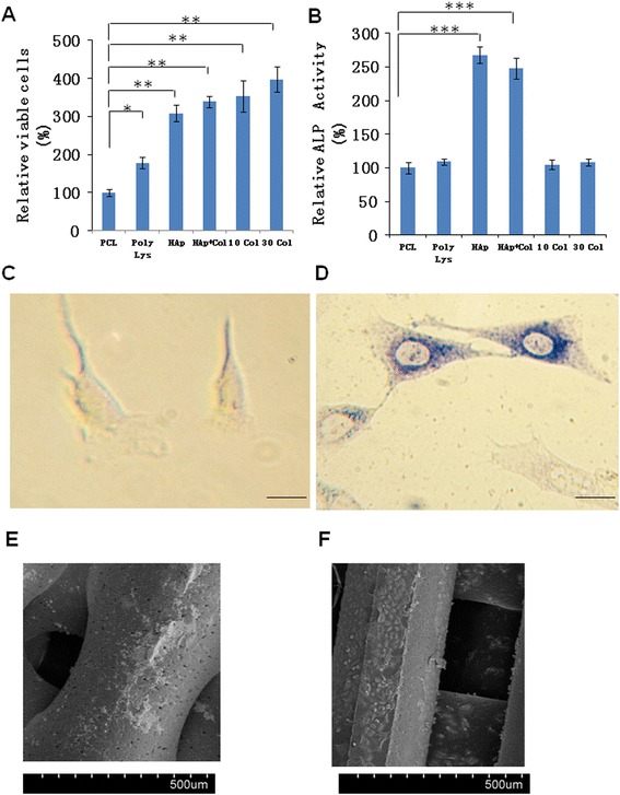
Results: Scanning Electron Microscope (SEM) micrographs showed that coating materials were uniformly deposited on the surface of PCL scaffolds and BMSCs grew and aggregated to form clusters during 3D culture. Both mRNA and protein levels of the key players of osteogenesis and osteoblast differentiation and activation, including runt-related transcription factor 2 (Runx2), alkaline phosphates (ALP), osterix, osteocalcin, and RANKL, were significantly higher in BMSCs seeded in PCL scaffolds coated with HAp or HAp/collagen than those seeded in uncoated PCL scaffolds, whereas the expression levels were not significantly different in collagen or poly-lysine coated PCL scaffolds. In addition, poly-lysine, collagen, HAp/collagen, and HAp coated PCL scaffolds had significantly more viable cells than uncoated PCL scaffolds, especially scaffolds with HAp/collagen and collagen-alone coatings. That BMSCs in HAp or HAp/collagen PCL scaffolds had remarkably higher ALP activities than those in collagen-coated alone or uncoated PCL scaffolds indicating higher osteogenic differentiation levels of BMSCs in HAp or HAp/collagen PCL scaffolds. Moreover, morphological changes of BMSCs after four-week of 3D culture confirmed that BMSCs successfully differentiated into osteoblast with spread-out phenotype in HAp/collagen coated PCL scaffolds.

Conclusion: This study showed a proof of concept for preparing biomimetic 3D poly (ε-caprolactone)/ hydroxyapatite/collagen scaffolds with excellent osteoinduction and proliferation capacity for bone regeneration.


Figures

Figure 1
Figure 1
Representative SEM micrographs of PCL scaffolds after coating. The micrographs show the details of scaffolds after coating. A, PCL scaffolds only; B, PCL scaffolds with poly-lysine coating; C, PCL scaffolds with HAp; D, PCL scaffolds with HAp and 30 mg/ml collagen coating; E, PCL scaffolds with 10 mg/ml collagen coating; F, PCL scaffolds with 30 mg/ml collagen coating.
Figure 2
Figure 2
Representative SEM micrographs of PCL scaffolds after coating and 28 days of culturing. The micrographs show the proliferation and spreading of BMSCs (white arrows) after 28 days in culture. A, PCL scaffolds only; B, PCL scaffolds with poly-lysine coating; C, PCL scaffolds with HAp; D, PCL scaffolds with HAp and 30 mg/ml collagen coating; E, PCL scaffolds with 10 mg/ml collagen coating; F, PCL scaffolds with 30 mg/ml collagen coating.
Figure 3
Figure 3
Osteogenic differentiation of BMSCs growing on scaffolds. The mRNA expression of bone tissue specific markers, including Runx2 (A), ALP (B), Osterix (C), Osteocalcin (D), RANKL (E) were evaluated during the in vitro culturing period of up to 28 days to assess osteogenic differentiation of BMSCs by RT-qPCR at different time points. All data were normalized to the mRNA expression of the corresponding marker at day 0 (cell seeding day). Data indicate the mean relative values calculated from six independent experiments (±S.E.). Statistically significant differences with P < 0.05 were considered significant (*P < 0.05). F, shows a representative protein expression (as indicated) of cell lysates cultured on PCL scaffolds only, PCL scaffolds with poly-lysine coating, PCL scaffolds with HAp, PCL scaffolds with HAp and 30 mg/ml collagen coating, PCL scaffolds with 10 mg/ml collagen coating, PCL scaffolds with 30 mg/ml collagen coating after 28 days of cell culturing.
Figure 4
Figure 4
Cell-scaffold interactions on PCL scaffolds. BMSCs were cultured for 4 weeks in vitro. A, cell proliferation of BMSCs on PCL scaffolds with or without coatings as indicated. Cell proliferation were determined via MTT assays. Viability of cells were normalized to the viability on PCL scaffolds only. B, alkaline phosphates (ALP) activity after 4 weeks of BMSC culturing. Data were normalized to the ALP activity on PCL scaffolds only. Data indicate the mean relative values calculated from six independent experiments (±S.E.). Statistically significant differences with P < 0.05 were considered significant (*P < 0.05; **P < 0.01; ***P < 0.001). C, representative BMSC image before culturing on PCL scaffolds with HAp and 30 mg/ml collagen coating. Scale bar, 10 μm. D, representative cell image after culturing on PCL scaffolds with HAp and 30 mg/ml collagen coating. The cells have differentiated into osteoblastic cells and were stained with ALP staining kit. SEM micrographs of PCL scaffolds after coating and 4 weeks of culturing. Scale bar, 10 μm. E, PCL scaffolds only. F, PCL scaffolds with HAp and 30 mg/ml collagen coating.

References

    1. Nudelman F, Lausch AJ, Sommerdijk NA, Sone ED. In vitro models of collagen biomineralization. J Struct Biol. 2013;183(2):258–69. doi: 10.1016/j.jsb.2013.04.003. - DOI - PubMed
    1. Dorozhkin SV. Calcium orthophosphates: occurrence, properties, biomineralization, pathological calcification and biomimetic applications. Biomatter. 2011;1(2):121–64. doi: 10.4161/biom.18790. - DOI - PMC - PubMed
    1. Daculsi G, Bouler JM, LeGeros RZ. Adaptive crystal formation in normal and pathological calcifications in synthetic calcium phosphate and related biomaterials. Int Rev Cytol. 1997;172:129–91. doi: 10.1016/S0074-7696(08)62360-8. - DOI - PubMed
    1. Boskey AL, Roy R. Cell culture systems for studies of bone and tooth mineralization. Chem Rev. 2008;108(11):4716–33. doi: 10.1021/cr0782473. - DOI - PMC - PubMed
    1. Meneghini C, Dalconi MC, Nuzzo S, Mobilio S, Wenk RH. Rietveld refinement on x-ray diffraction patterns of bioapatite in human fetal bones. Biophys J. 2003;84(3):2021–9. doi: 10.1016/S0006-3495(03)75010-3. - DOI - PMC - PubMed

Publication types

MeSH terms

LinkOut - more resources