

Clinical characteristics of responders to intravitreal bevacizumab in central serous chorioretinopathy patients
- PMID: 25952951
- PMCID: PMC4469674
- DOI: 10.1038/eye.2015.58
Clinical characteristics of responders to intravitreal bevacizumab in central serous chorioretinopathy patients
Abstract
Purpose: To investigate factors associated with good response to intravitreal bevacizumab (IVB) in central serous chorioretinopathy (CSC) patients.
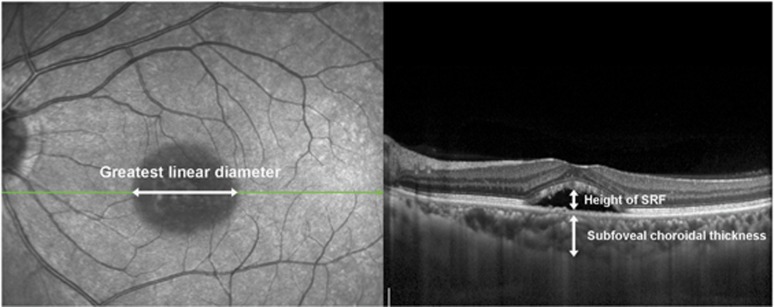
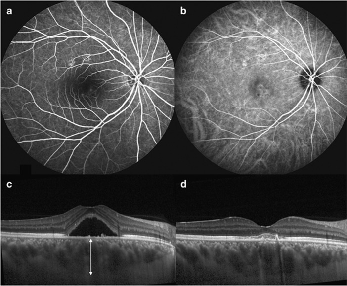
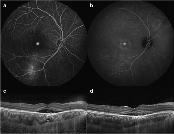
Methods: We retrospectively reviewed 42 eyes of CSC patients of symptom duration more than 3 months who received a single or multiple successive IVBs on an as-needed basis (0.05 ml, 1.25 mg). High responders (HRs) were defined as complete resolution of subretinal fluid (SRF) on spectral domain optical coherence tomography (SD-OCT). Moderate responders (MRs) were defined as SRF resolution of 50-99% of pretreatment volume and poor responders (PRs) as SRF resolution <50%. Clinical, SD-OCT, fluorescein, and indocyanine green angiography findings were analyzed to find factors associated with HR. Descriptive statistics for all demographic and clinical variables were calculated, and comparisons were made using Wilcoxon's matched-pairs signed-rank test, the Mann-Whitney U-test for means with continuous data, Pearson's χ(2) test, and Fisher's exact test for categorical data.
Results: The mean number of IVB was 1.9. At postoperative 1 month, there were 10 (24%) HRs, 18 (43%) MRs, and 14 (33%) PRs. At the last follow-up (the mean 8.6 months), there were 25 HRs (60%), 9 MRs (21%), and 8 PRs (19%). Thicker subfoveal choroid (P=0.036), smaller lesion diameter (P=0.019), and better baseline best-corrected visual acuity (P=0.002) predicted HRs at postoperative 1 month. HR at the last follow-up was associated with classic pattern fluorescein angiography finding.
Conclusions: Suboptimal effects of IVB on persistent CSC suggest primary IVB on selective cases with better vision, smaller lesion, and thicker choroid at baseline.
Figures
Similar articles
-
SUBFOVEAL CHOROIDAL THICKNESS CHANGES IN TREATED IDIOPATHIC CENTRAL SEROUS CHORIORETINOPATHY AND THEIR ASSOCIATION WITH RECURRENCE.Retina. 2015 Sep;35(9):1867-74. doi: 10.1097/IAE.0000000000000557. Retina. 2015. PMID: 25946693
-
FLAT IRREGULAR PIGMENT EPITHELIUM DETACHMENT IN CENTRAL SEROUS CHORIORETINOPATHY: A Form of Pachychoroid Neovasculopathy?Retina. 2020 Sep;40(9):1724-1733. doi: 10.1097/IAE.0000000000002662. Retina. 2020. PMID: 31584559
-
Intravitreal Bevacizumab and Ranibizumab in the Treatment of Acute Central Serous Chorioretihopathy: A Single Center Retrospective Study.Semin Ophthalmol. 2018;33(2):265-270. doi: 10.1080/08820538.2016.1228985. Epub 2016 Nov 14. Semin Ophthalmol. 2018. PMID: 27841949
-
Lack of positive effect of intravitreal bevacizumab in central serous chorioretinopathy: meta-analysis and review.Eye (Lond). 2013 Dec;27(12):1339-46. doi: 10.1038/eye.2013.236. Epub 2013 Nov 8. Eye (Lond). 2013. PMID: 24202051 Free PMC article. Review.
-
Clinical efficacy of anti-VEGF medications for central serous chorioretinopathy: a meta-analysis.Int J Clin Pharm. 2017 Jun;39(3):514-521. doi: 10.1007/s11096-017-0460-4. Epub 2017 Apr 6. Int J Clin Pharm. 2017. PMID: 28386700 Review.
Cited by
-
Therapeutic Efficacy of Spironolactone for Central Serous Chorioretinopathy.Yonsei Med J. 2022 Apr;63(4):365-371. doi: 10.3349/ymj.2022.63.4.365. Yonsei Med J. 2022. PMID: 35352888 Free PMC article.
-
Photodynamic therapy in combination with intravitreal ziv-aflibercept and aflibercept injection in patients with chronic or repeatedly recurrent acute central serous chorioretinopathy: a single-center retrospective study.Clin Ophthalmol. 2018 Jul 20;12:1301-1309. doi: 10.2147/OPTH.S165199. eCollection 2018. Clin Ophthalmol. 2018. PMID: 30050283 Free PMC article.
-
Visual and Anatomical Outcomes of Spironolactone Therapy in Patients with Chronic Central Serous Chorioretinopathy.J Ophthalmic Vis Res. 2017 Jul-Sep;12(3):281-289. doi: 10.4103/jovr.jovr_139_16. J Ophthalmic Vis Res. 2017. PMID: 28791061 Free PMC article.
-
Long-term treatment response after intravitreal bevacizumab injections for patients with central serous chorioretinopathy.PLoS One. 2020 Sep 8;15(9):e0238725. doi: 10.1371/journal.pone.0238725. eCollection 2020. PLoS One. 2020. PMID: 32898167 Free PMC article.
-
Short-term effect of anti-VEGF for chronic central serous chorioretinopathy according to the presence of choroidal neovascularization using optical coherence tomography angiography.PLoS One. 2021 Jan 11;16(1):e0245342. doi: 10.1371/journal.pone.0245342. eCollection 2021. PLoS One. 2021. PMID: 33428683 Free PMC article.
References
-
- Wang M, Munch IC, Hasler PW, Prunte C, Larsen M. Central serous chorioretinopathy. Acta Ophthalmol. 2008;86 (2:126–145. - PubMed
-
- Prunte C. Indocyanine green angiographic findings in central serous chorioretinopathy. Int Ophthalmol. 1995;19 (2:77–82. - PubMed
-
- Prunte C, Flammer J. Choroidal capillary and venous congestion in central serous chorioretinopathy. Am J Ophthalmol. 1996;121 (1:26–34. - PubMed
-
- Hayashi K, Hasegawa Y, Tokoro T. Indocyanine green angiography of central serous chorioretinopathy. Int J Ophthalmol. 1986;9 (1:37–41. - PubMed
-
- Yannuzzi LA. Central serous chorioretinopathy: a personal perspective. Am J Ophthalmol. 2010;149 (3:361–363. - PubMed
MeSH terms
Substances
LinkOut - more resources
Full Text Sources
Other Literature Sources
Miscellaneous
