. 2015;10(13):2051-61.
doi: 10.2217/nnm.15.52. Epub 2015 May 8.

Magnetoelectric 'spin' on stimulating the brain

Affiliations

Magnetoelectric 'spin' on stimulating the brain

Rakesh Guduru et al. Nanomedicine (Lond). 2015.

Abstract

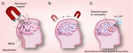
Aim: The in vivo study on imprinting control region mice aims to show that magnetoelectric nanoparticles may directly couple the intrinsic neural activity-induced electric fields with external magnetic fields.

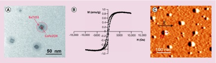
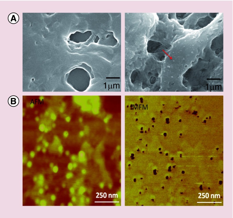
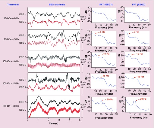
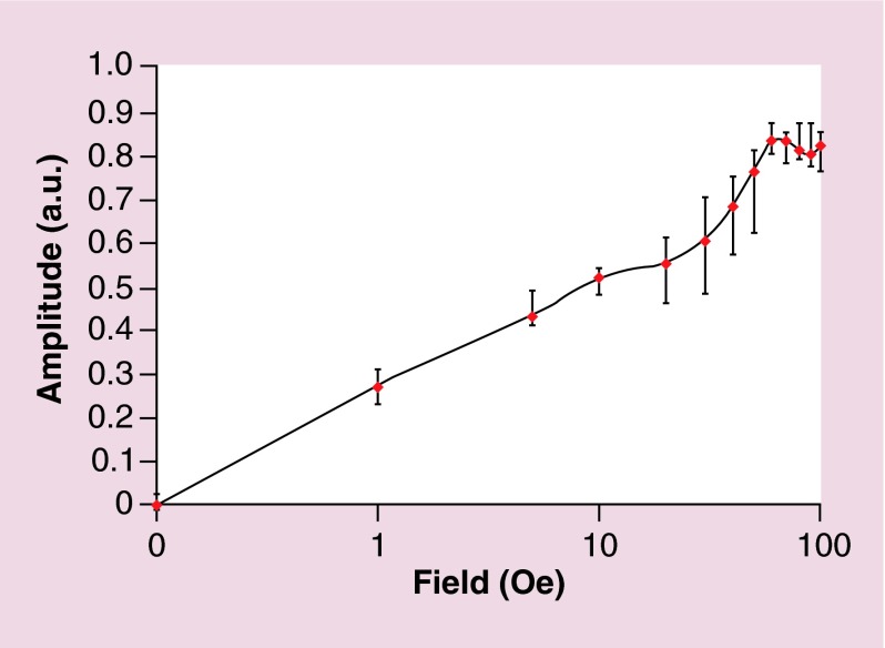
Methods: Approximately 10 µg of CoFe2O4-BaTiO3 30-nm nanoparticles have been intravenously administrated through a tail vein and forced to cross the blood-brain barrier via a d.c. field gradient of 3000 Oe/cm. A surgically attached two-channel electroencephalography headmount has directly measured the modulation of intrinsic electric waveforms by an external a.c. 100-Oe magnetic field in a frequency range of 0-20 Hz.

Results: The modulated signal has reached the strength comparable to that due the regular neural activity.

Conclusion: The study opens a pathway to use multifunctional nanoparticles to control intrinsic fields deep in the brain.

Keywords: magnetoelectric nanoparticles; nanoengineering the brain; noninvasive brain stimulation.


Conflict of interest statement

Financial & competing interests disclosure The authors acknowledge partial financial support from National Science Foundation (NSF) awards #ECCS-1408063 and IIP-1237818, NIH DA #R01DA034547-01 and Department of Defense (DoD) Defense Microelectronics Activity (DMEA) under contract #H94003–09–2–0904. The authors have no other relevant affiliations or financial involvement with any organization or entity with a financial interest in or financial conflict with the subject matter or materials discussed in the manuscript apart from those disclosed. No writing assistance was utilized in the production of this manuscript.

Figures

<b>Figure 1.</b>
Figure 1.. Nanoparticle characterization.
(A) Transmission electron microscopy image of magnetoelectric nanoparticles (MENs). The core-shell structure can be seen. (B) M-H loop of MENs measured via vibrating sample magnetometry at room temperature. (C) Magnetic force microscopy image of MENs exposed to an external electric field Ex of 100 V/cm. M stands for the magnetization.
<b>Figure 2.</b>
Figure 2.. Detection of magnetoelectric nanoparticles deep in the brain.
(A) Scanning electron microscopy images of sagittal brain sections of two mice: (left) without magnetoelectric nanoparticles (MENs) in the brain and (right) with MENs in the brain. The red arrow points to a MEN. (B) Atomic force microscopy (left) and magnetic force microscopy images of a small region containing a relatively dense concentration of MENs. AFM: Atomic force microscopy; MFM: Magnetic force microscopy.
<b>Figure 3.</b>
Figure 3.. Key measurements – EEG waveforms with magnetoelectric nanoparticles in the brain.
EEG Waveforms (left) and their frequency spectra (right) from the two EEG channels with MENs in the brain under exposure to an external 100-Oe a.c. magnetic field at frequencies of 0, 5, 10, 15 and 20 Hz. The vertical scale bar for the waveform signal is 5 mV.
<b>Figure 4.</b>
Figure 4.. a.c. field dependence of the modulation effect.
The modulation amplitude of the electric signal (in arbitrary units) versus the external a.c. magnetic field amplitude. The field axis is shown in a logarithmic scale.
<b>Figure 5.</b>
Figure 5.. Concept illustration.
Schematic illustration of the novel concept to use MENs for ‘mapping’ the brain for noninvasive electric field stimulation of selected regions deep in the brain. (A) MENs are forced into the brain across blood–brain barrier via application of a d.c. magnetic field gradient. (B) When in the brain, MENs are being distributed over the entire brain or in selected regions via application of spatially varying d.c. magnetic field gradients. The presence of MENs effectively creates a ‘new brain microenvironment’, in which the intrinsic electric signals due to the neural activity are strongly coupled at the subneuronal level to the external magnetic fields generated by remote sources. (C) Such coupling can be used for noninvasive high-efficacy stimulation of selected regions deep in the brain via application of focused and relatively low (˜100 Oe) near-d.c. (˜<1000 Hz) magnetic field. MEN: Magnetoelectric nanoparticle.

References

    1. Marblestone AH, et al. Physical principles for scalable neural recording. Front. Comput. Neurosci. 2013;7:137. - PMC - PubMed
    2. • Gives a unique technology angle on the current state of neuroengineering.

    1. Hopfield JJ. Neurons with graded response have collective computational properties like those of two-state neurons. Proc. Natl Acad. Sci. USA. 1984;81:3088–3092. - PMC - PubMed
    1. De Kramer J. The electrical circuitry of an olfactory sensillum in Antheraea polyphemus . J. Neurosci. 1985;5:2484–2493. - PMC - PubMed
    2. • Presents the concept of the brain's electric circuitry.

    1. Srikanth M, Kessler JA. Nanotechnology – novel therapeutics for central nervous system disorders. Nat. Rev. Neurol. 2012;8:307–318. - PMC - PubMed
    2. • Gives an overview of using nanotechnology for treating CNS disorders.

    1. Yue K, et al. Magneto-electric nano-particles for non-invasive brain stimulation. PLoS ONE. 2012;7:e44040. - PMC - PubMed
    2. • Gives a theoretical insight into using magnetoelectric nanoparticles for noninvasive brain stimulation.

Publication types

MeSH terms