. 2015 Jul;138(Pt 7):1907-18.
doi: 10.1093/brain/awv107. Epub 2015 May 6.

The role of tau in the pathological process and clinical expression of Huntington's disease

Affiliations

The role of tau in the pathological process and clinical expression of Huntington's disease

Romina Vuono et al. Brain. 2015 Jul.

Abstract

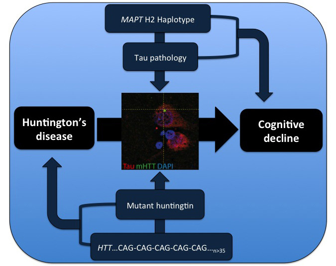
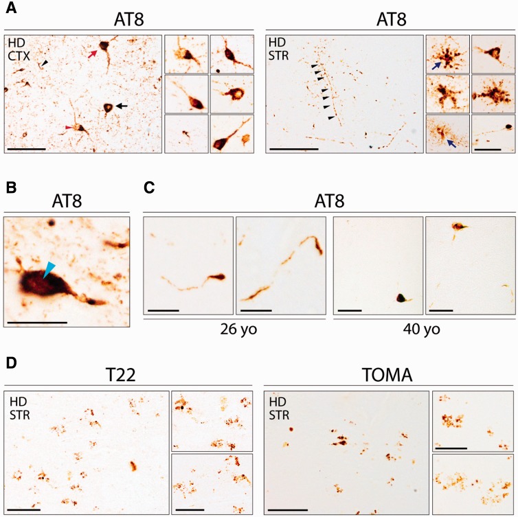
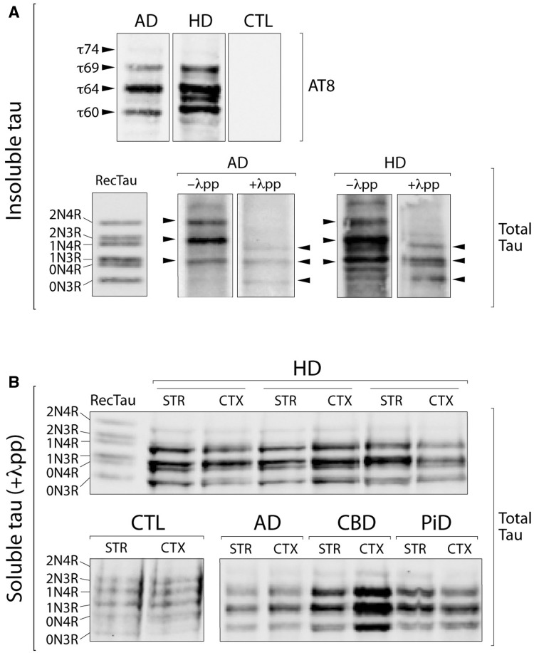
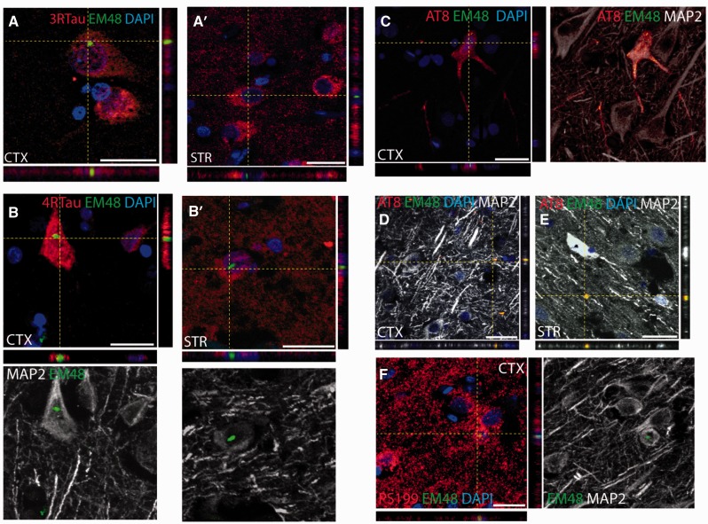
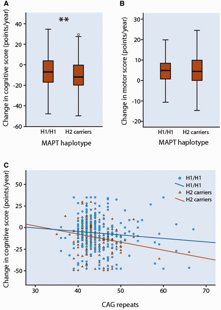
Huntington's disease is a neurodegenerative disorder caused by an abnormal CAG repeat expansion within exon 1 of the huntingtin gene HTT. While several genetic modifiers, distinct from the Huntington's disease locus itself, have been identified as being linked to the clinical expression and progression of Huntington's disease, the exact molecular mechanisms driving its pathogenic cascade and clinical features, especially the dementia, are not fully understood. Recently the microtubule associated protein tau, MAPT, which is associated with several neurodegenerative disorders, has been implicated in Huntington's disease. We explored this association in more detail at the neuropathological, genetic and clinical level. We first investigated tau pathology by looking for the presence of hyperphosphorylated tau aggregates, co-localization of tau with mutant HTT and its oligomeric intermediates in post-mortem brain samples from patients with Huntington's disease (n = 16) compared to cases with a known tauopathy and healthy controls. Next, we undertook a genotype-phenotype analysis of a large cohort of patients with Huntington's disease (n = 960) with a particular focus on cognitive decline. We report not only on the tau pathology in the Huntington's disease brain but also the association between genetic variation in tau gene and the clinical expression and progression of the disease. We found extensive pathological inclusions containing abnormally phosphorylated tau protein that co-localized in some instances with mutant HTT. We confirmed this related to the disease process rather than age, by showing it is also present in two patients with young-onset Huntington's disease (26 and 40 years old at death). In addition we demonstrate that tau oligomers (suggested to be the most likely neurotoxic tau entity) are present in the Huntington's disease brains. Finally we highlight the clinical significance of this pathology by demonstrating that the MAPT haplotypes affect the rate of cognitive decline in a large cohort of patients with Huntington's disease. Our findings therefore highlight a novel important role of tau in the pathogenic process and clinical expression of Huntington's disease, which in turn opens up new therapeutic avenues for this incurable condition.

Keywords: Huntington’s disease; dementia; neurofibrillary tangles; neuropathology; tau.


Figures

None
Tau has recently been implicated in Huntington’s disease, but the nature of its involvement is unclear. Vuono et al. reveal tau oligomers and hyperphosphorylated tau aggregates in post-mortem Huntington’s disease brains, including those from young-onset cases. Genotype-phenotype analysis of a large patient cohort shows that tau haplotypes influence cognitive decline.
Figure 1
Figure 1
Tau pathology in Huntington’s disease brains. (A) Immunohistochemical detection of pathological tau aggregates in cortical (CTX) and striatal (STR) Huntington’s disease tissue [HD8, female, 61 years old (yo)] using the AT8 antibody against phosphorylated tau. Neuronal inclusions including ring-like perinuclear (black arrow), flame shaped (red arrow), and globular (red arrowhead) morphologies were observed. Tufted astrocytes and astrocytic plaques (blue arrows) numerous dots and neuropil threads (black arrowhead) were also seen. Scale bars = 50 μm, insets = 20 μm. (B) Nuclear rod-like tau deposits positive for AT8-immunoreactivity in Huntington’s disease brains (light blue arrowhead). Scale bar = 20 µm. (C) Phosphorylated tau aggregates detected by immunohistochemistry using AT8 in both cases of patients with young onset Huntington’s disease (26 and 40 years old). Scale bars = 10 µm (26-years-old) and 50 µm (40-years-old). (D) Immunohistochemistry using TOMA and T22 antibodies in striatal Huntington’s disease tissue (HD8, female, 61-years-old) showing oligomeric tau inclusions. Scale bars = 100 μm, insets = 10 μm.
Figure 2
Figure 2
Biochemical characterization of sarkosyl-insoluble and soluble tau in Huntington’s disease brains. (A) Western blot analysis of the sarkosyl-insoluble tau fraction, before and after dephosphorylation with alkaline phosphatase (λpp), with the AT8 and a total tau antibody in cortical and striatal Huntington’s disease tissues compared to Alzheimer’s disease (AD) cases and healthy controls (CTL). (B) Western blot analysis of the soluble tau fraction after dephosphorylation with alkaline phosphatase (λpp) with a total tau antibody in cortical (CTX) and striatal (STR) Huntington’s disease tissues, compared to a range of tauopathies (CBD = corticobasal degeneration; PiD = Pick’s disease) and healthy controls. RecTau molecular weight (kDa): 2N4R (45.9), 2N3R (42.6), 1N4R (42.9), 1N3R (39.7), 0N4R (40.0), 0N3R (36.8).
Figure 3
Figure 3
Mutant huntingtin co-localizes with pathological tau aggregates. (A and B) Mutant HTT aggregates labelled with the EM48 antibody (green) detected within cortical and striatal neurons expressing either 3R-tau or 4R-tau (red). Scale bars in A, A’, B = 25 μm; B’ = 50 μm. (C–F) Confocal microscopy images of mutant HTT aggregates labelled with the EM48 antibody (green) within neurons expressing phosphorylated tau (red) as demonstrated using either the AT8 or pS199 antibodies. The neuronal morphology was further confirmed using the neuronal marker MAP2 (grey). Aggregates stained for both EM48 and AT8 were also found in the extracellular matrix of the cortex and striatum of Huntington’s disease cases as shown in D and E. Scale bars in C–E = 30 μm; F = 20 μm.
Figure 4
Figure 4
The effect of MAPT haplotypes on cognitive and motor decline in patients with Huntington’s disease. (A) Box and whisker plot showing rates of change in composite cognitive scores in patients with Huntington’s disease grouped by MAPT haplotype. Median, interquartile range, and minimum-maximum range are shown. Distributions were compared using Mann–Whitney U-tests. **P < 0.01. (B) Box and whisker plot showing rates of change in UHDRS Motor scores in patients with Huntingtons' disease grouped by MAPT haplotype. (C) Correlation between the CAG repeat length and change in composite cognitive score per year in patients with Huntington’s disease grouped by MAPT haplotype. Across all participants (n = 473), Kendall’s taub = −0.098, P = 0.002. In H1/H1 (squares; n = 283), Kendall’s taub = −0.063, P = 0.129; in H2 carriers (triangles; n = 190); Kendall’s taub = −0.148, P = 0.004.

Comment in

References

    1. Anfossi M, Vuono R, Maletta R, Virdee K, Mirabelli M, Colao R, et al. Compound heterozygosity of 2 novel MAPT mutations in frontotemporal dementia. Neurobiol Aging. 2011;32:757, e1–e11. - PubMed
    1. Baker M, Litvan I, Houlden H, Adamson J, Dickson D, Perez-Tur J, et al. Association of an extended haplotype in the tau gene with progressive supranuclear palsy. Hum Mol Genet. 1999;8:711–15. - PubMed
    1. Blum D, Herrera F, Francelle L, Mendes T, Basquin M, Obriot H, et al. Mutant huntingtin alters Tau phosphorylation and subcellular distribution. Hum Mol Genet. 2015;24:76–85. - PubMed
    1. Braak H, Braak E. Neuropathological stageing of Alzheimer-related changes. Acta Neuropathol. 1991;82:239–59. - PubMed
    1. Castillo-Carranza DL, Sengupta U, Guerrero-Muñoz MJ, Lasagna-Reeves CA, Gerson JE, Singh G, et al. Passive immunization with Tau oligomer monoclonal antibody reverses tauopathy phenotypes without affecting hyperphosphorylated neurofibrillary tangles. J Neurosci. 2014;34:4260–72. - PMC - PubMed

Publication types