. 2015 Aug;29(8):3527-36.
doi: 10.1096/fj.15-271304. Epub 2015 May 7.

Alternative activation of macrophages and pulmonary fibrosis are modulated by scavenger receptor, macrophage receptor with collagenous structure

Affiliations

Alternative activation of macrophages and pulmonary fibrosis are modulated by scavenger receptor, macrophage receptor with collagenous structure

Shubha Murthy et al. FASEB J. 2015 Aug.

Abstract

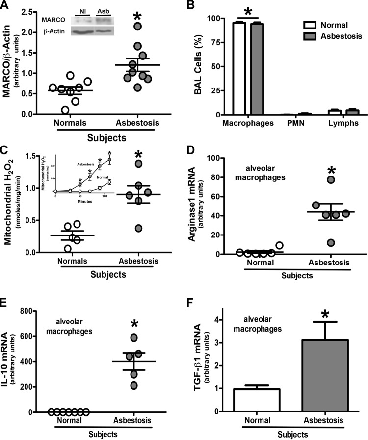
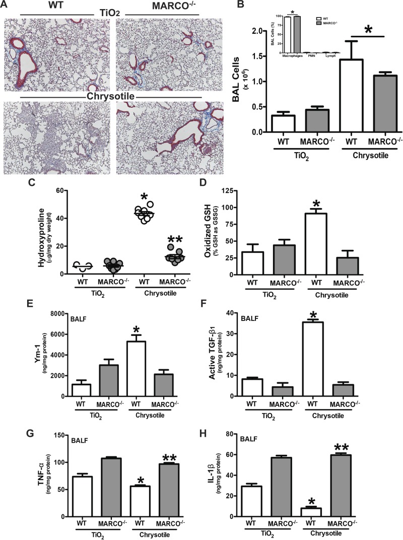
Alternative activation of alveolar macrophages is linked to fibrosis following exposure to asbestos. The scavenger receptor, macrophage receptor with collagenous structure (MARCO), provides innate immune defense against inhaled particles and pathogens; however, a receptor for asbestos has not been identified. We hypothesized that MARCO acts as an initial signaling receptor for asbestos, polarizes macrophages to a profibrotic M2 phenotype, and is required for the development of asbestos-induced fibrosis. Compared with normal subjects, alveolar macrophages isolated from patients with asbestosis express higher amounts of MARCO and have greater profibrotic polarization. Arginase 1 (40-fold) and IL-10 (265-fold) were higher in patients. In vivo, the genetic deletion of MARCO attenuated the profibrotic environment and pulmonary fibrosis in mice exposed to chrysotile. Moreover, alveolar macrophages from MARCO(-/-) mice polarize to an M1 phenotype, whereas wild-type mice have higher Ym1 (>3.0-fold) and nearly 7-fold more active TGF-β1 in bronchoalveolar lavage (BAL) fluid (BALF). Arg(432) and Arg(434) in domain V of MARCO are required for the polarization of macrophages to a profibrotic phenotype as mutation of these residues reduced FIZZ1 expression (17-fold) compared with cells expressing MARCO. These observations demonstrate that a macrophage membrane protein regulates the fibrotic response to lung injury and suggest a novel target for therapeutic intervention.

Keywords: MARCO; asbestos; mitochondria.


Figures

Figure 1.
Figure 1.
Alveolar macrophages from patients with asbestosis have higher MARCO expression, greater mitochondrial oxidative stress, and a predominant M2 phenotype. A) MARCO and β-actin expression in alveolar macrophages obtained by BAL from normal subjects (Nl, n = 8) and patients with asbestosis (Asb, n = 9) were determined by immunoblotting. Densitometric analysis and a representative blot (inset) are shown. Results are expressed as arbitrary units. *P < 0.05. B) Cells were obtained by BAL from normal subjects (n = 5) and patients with asbestosis (n = 5). Cell differential was determined by Wright-Giemsa stain. *P < 0.0001 macrophages vs. all other cell types. Mitochondria were isolated from alveolar macrophages from normal subjects and patients with asbestosis. C) H2O2 production rate and (inset) H2O2 production measured by pHPA assay. Results are expressed as nanomoles per milligram per minute. *P < 0.05. Nl (n = 5) and Asb (n = 6). Total RNA was isolated from alveolar macrophages obtained from normal subjects and patients with asbestosis by BAL. D) Arginase 1, *P < 0.0008 (n = 6); (E) IL-10, *P < 0.0001 (Nl, n = 7; Asb, n = 5); and (F) TGF-β1 mRNA, * P < 0.03 (n = 5) were measured and are expressed in arbitrary units.
Figure 2.
Figure 2.
Asbestos induces MARCO expression in vivo and in vitro. A) WT mice were intratracheally administered 100 μg/mouse chrysotile asbestos and, at the indicated times, mice were killed and alveolar macrophages were obtained by BAL. Cells were lysed, and MARCO and β-actin expression determined by immunoblotting. Alveolar macrophages were exposed to (B) chrysotile or (C) TiO2 for the designated time, and MARCO, SR-A, and β-actin expression were determined by immunoblot analysis. D) Macrophages were exposed to chrysotile for 180 min. Membrane and cytosol fractions were isolated and MARCO, gp91phox, and β-actin expression were determined by immunoblot analysis.
Figure 3.
Figure 3.
MARCO is required for the development of pulmonary fibrosis. WT and MARCO−/− mice were intratracheally administered 100 μg/mouse of TiO2 or chrysotile asbestos. Twenty-one days later, mice were killed, BAL was performed, and lungs were removed. A) Lungs were stained for collagen with Masson’s trichrome stain. Representative sections from WT (n = 4) and MARCO−/− (n = 4) mice are shown. B) Cells obtained by BAL from WT (n = 5, TiO2; n = 6, chrysotile) and MARCO−/− (n = 5, TiO2; n = 7, chrysotile) mice. *P < 0.0001 vs. TiO2-exposed mice. (Inset) Cell differential from chrysotile-exposed mice was determined by Wright-Giemsa stain. *P < 0.0001 (n = 4) macrophages vs. all other cell types. C) Hydroxyproline content in lungs is shown as microgram hydroxyproline per milligram lung dry weight. *P < 0.0001 vs. all other groups, and **P < 0.0014 vs. TiO2-exposed mice. WT (n = 3, TiO2; n = 8, chrysotile) and MARCO−/− (n = 8, TiO2; n = 7, chrysotile) mice. D) GSH and GSSG were measured by HPLC and mass spectrometry analysis and are expressed as oxidized glutathione (GSSG) relative to its reduced form (GSH). *P < 0.05. WT (n = 6) and MARCO−/− (n = 7, TiO2; n = 6, chrysotile) mice. BALF was obtained by BAL. ELISA was performed for (E) Ym-1, *P < 0.0002 vs. all other groups, WT (n = 5, TiO2; n = 8, chrysotile) and MARCO−/− (n = 7, TiO2; n = 8, chrysotile); (F) active TGF-β1, *P < 0.0001 vs. all other groups; (G) TNF-α, *P < 0.0001 vs. all other groups and **P < 0.0009 vs. WT groups; and (H) IL-1β, * P < 0.0001 vs. all other groups and **P < 0.0001 vs. WT groups. WT (n = 4, TiO2; n = 8, chrysotile) and MARCO−/− (n = 5, TiO2; n = 8, chrysotile) (F–H).
Figure 4.
Figure 4.
MARCO is required for mitochondrial H2O2 production. A) Macrophages were incubated for 1 h with 500 μg/ml fucoidan before being exposed to chrysotile. Mitochondria were isolated and the production of H2O2 was determined. Results are expressed as nanomoles per milligram per minute. *P < 0.0001 control + chrysotile vs. fucoidan + chrysotile. n = 3. B) Macrophages were transfected with either scrambled or MARCO siRNA, and 48 h later cells were cultured in the presence or absence of chrysotile. Mitochondria were isolated and H2O2 production was measured. Results are expressed as nanomoles per milligram per minute. *P < 0.0001 vs. all other groups. n = 3. (Inset) Knockdown of MARCO by immunoblot analysis. C) Macrophages were transfected as in B. After 48 h, cells were exposed to chrysotile. Cell binding to chrysotile was counted in fifteen fields by microscopy. n = 15. * P < 0.0004. (Inset) Knockdown of MARCO by immunoblot analysis. D) Cells were transfected with scrambled or Rieske siRNA and 48 h later transfected with empty pcDNA3.1 or vector expressing MARCOWT. Twenty-four hours later, cells were exposed to chrysotile and H2O2 production was determined in whole cells. Results are expressed as nanomoles per milligram per minute. n = 3. *P < 0.05 –chrysotile vs. +chrysotile; **P < 0.05 vs. scrambled siRNA + empty + chrysotile; #P < 0.05 vs. scrambled siRNA + empty + chrysotile; ##P < 0.05 vs. scrambled siRNA + MARCOWT + chrysotile. (Inset) Rieske knockdown was determined by immunoblotting.
Figure 5.
Figure 5.
Arginine residues in domain V of MARCO mediates mitochondrial ROS and promotes M2 polarization. A) Schematic diagram of truncation mutants of MARCO. B) Macrophages were transfected with empty or vectors expressing MARCOWT or truncation mutants. After 24 h cells were exposed to chrysotile and H2O2 production was measured. C) Schematic diagram of site-directed MARCO mutants. D) Macrophages were transfected with empty or vectors expressing MARCOWT or site-directed mutants. After 24 h cells were exposed to chrysotile, mitochondria were isolated, and H2O2 production was measured. B, D) Results show mean ± sem, n = 3, *P < 0.05 –chrysotile vs. +chrysotile; **P < 0.05 vs. MARCOWT + chrysotile. Macrophages were transfected with empty vector or vectors expressing WT or R432A,R434A mutant and 24 h later were exposed to chrysotile for (E) 4 or (F) 24 h. E) Fizz-1 mRNA and F) arginase activity were determined. Results show mean ± sem, n = 3, *P < 0.05 –chrysotile vs. +chrysotile; **P < 0.05 vs. MARCOWT + chrysotile; ***P < 0.05 vs. empty + chrysotile and MARCOWT + chrysotile.

References

    1. Harraz M. M., Marden J. J., Zhou W., Zhang Y., Williams A., Sharov V. S., Nelson K., Luo M., Paulson H., Schöneich C., Engelhardt J. F. (2008) SOD1 mutations disrupt redox-sensitive Rac regulation of NADPH oxidase in a familial ALS model. J. Clin. Invest. 118, 659–670 - PMC - PubMed
    1. Gordon S., Martinez F. O. (2010) Alternative activation of macrophages: mechanism and functions. Immunity 32, 593–604 - PubMed
    1. Martinez F. O., Helming L., Gordon S. (2009) Alternative activation of macrophages: an immunologic functional perspective. Annu. Rev. Immunol. 27, 451–483 - PubMed
    1. Herbert D. R., Hölscher C., Mohrs M., Arendse B., Schwegmann A., Radwanska M., Leeto M., Kirsch R., Hall P., Mossmann H., Claussen B., Förster I., Brombacher F. (2004) Alternative macrophage activation is essential for survival during schistosomiasis and downmodulates T helper 1 responses and immunopathology. Immunity 20, 623–635 - PubMed
    1. Nair M. G., Du Y., Perrigoue J. G., Zaph C., Taylor J. J., Goldschmidt M., Swain G. P., Yancopoulos G. D., Valenzuela D. M., Murphy A., Karow M., Stevens S., Pearce E. J., Artis D. (2009) Alternatively activated macrophage-derived RELM-alpha is a negative regulator of type 2 inflammation in the lung. J. Exp. Med. 206, 937–952 - PMC - PubMed

Publication types