. 2015 Jun 26;290(26):16132-41.
doi: 10.1074/jbc.M115.656496. Epub 2015 May 7.

A Novel Mutation in Isoform 3 of the Plasma Membrane Ca2+ Pump Impairs Cellular Ca2+ Homeostasis in a Patient with Cerebellar Ataxia and Laminin Subunit 1α Mutations

Affiliations

A Novel Mutation in Isoform 3 of the Plasma Membrane Ca2+ Pump Impairs Cellular Ca2+ Homeostasis in a Patient with Cerebellar Ataxia and Laminin Subunit 1α Mutations

Tito Calì et al. J Biol Chem. .

Abstract

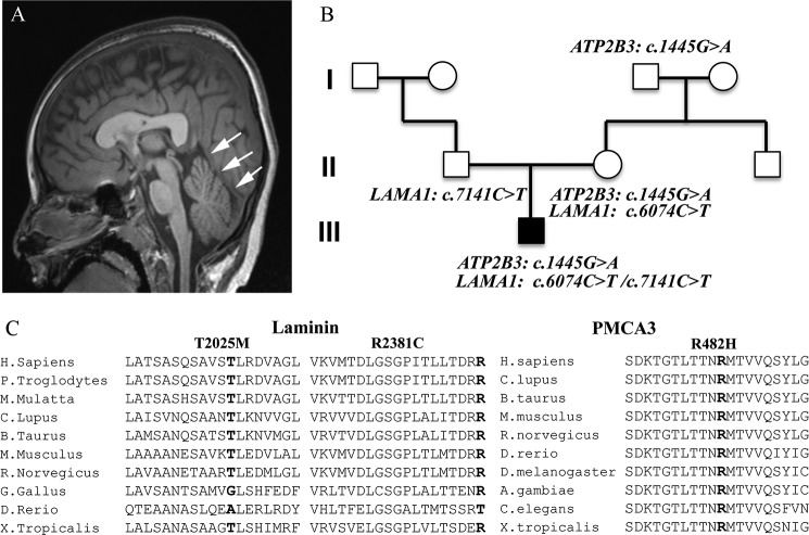
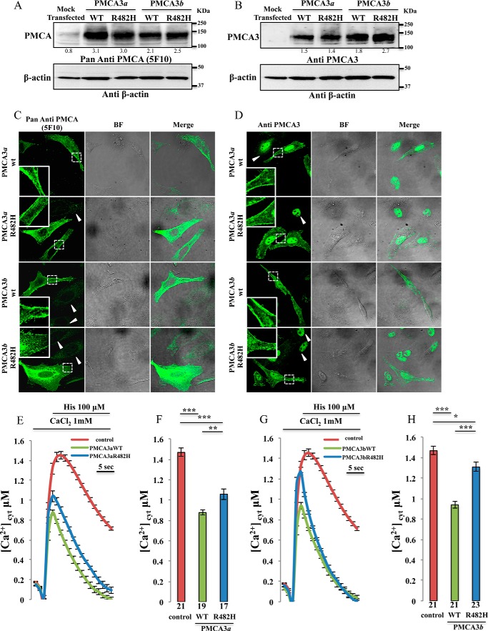
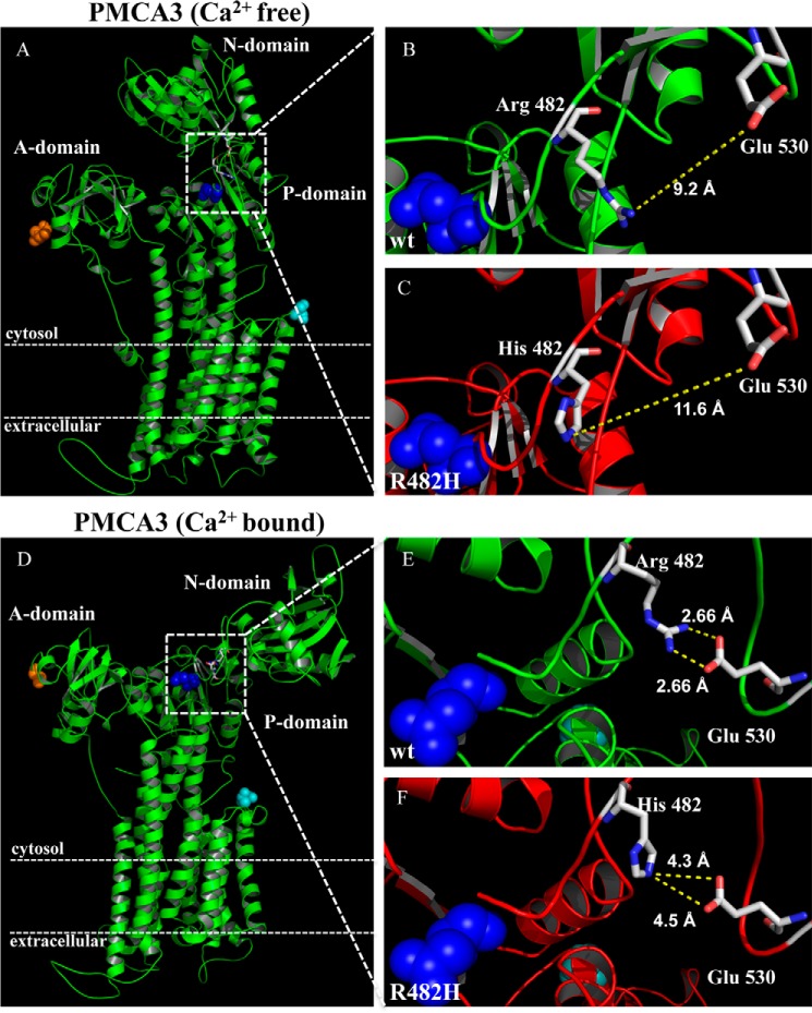
The particular importance of Ca(2+) signaling to neurons demands its precise regulation within their cytoplasm. Isoform 3 of the plasma membrane Ca(2+) ATPase (the PMCA3 pump), which is highly expressed in brain and cerebellum, plays an important role in the regulation of neuronal Ca(2+). A genetic defect of the PMCA3 pump has been described in one family with X-linked congenital cerebellar ataxia. Here we describe a novel mutation in the ATP2B3 gene in a patient with global developmental delay, generalized hypotonia and cerebellar ataxia. The mutation (a R482H replacement) impairs the Ca(2+) ejection function of the pump. It reduces the ability of the pump expressed in model cells to control Ca(2+) transients generated by cell stimulation and impairs its Ca(2+) extrusion function under conditions of low resting cytosolic Ca(2+) as well. In silico analysis of the structural effect of the mutation suggests a reduced stabilization of the portion of the pump surrounding the mutated residue in the Ca(2+)-bound state. The patient also carries two missense mutations in LAMA1, encoding laminin subunit 1α. On the basis of the family pedigree of the patient, the presence of both PMCA3 and laminin subunit 1α mutations appears to be necessary for the development of the disease. Considering the observed defect in cellular Ca(2+) homeostasis and the previous finding that PMCAs act as digenic modulators in Ca(2+)-linked pathologies, the PMCA3 dysfunction along with LAMA1 mutations could act synergistically to cause the neurological phenotype.

Keywords: ataxia; calcium; calcium ATPase; enzyme mutation; laminin.


Figures

FIGURE 1.
FIGURE 1.
Phenotype of the proband, pedigree family and multispecies alignment of the LAMA1 and PMCA3 mutations. A, brain imaging of the proband. Single T1-weighted midline sagittal image demonstrates unusual vertical course of the straight sinus (arrows) with inferior location of the torcula. This configuration is consistent with a small posterior fossa. B, pedigree of the proband's family showing the mapped mutations. C, multispecies alignment of the Laminin1 and the PMCA3 amino acids sequences showing that the mutated residues are conserved (bold).
FIGURE 2.
FIGURE 2.
Analysis of the expression of the exogenous pump variants and cytosolic Ca2+ measurements in HeLa cells overexpressing the wt and the mutant PMCA3 isoforms. A and B, Western blotting. C and D, immunocytochemistry analysis showing the expression level and the intracellular distribution of the PMCA3 pump in HeLa cells expressing the wt and the mutant truncated (a) as well as full-length (b) variants. The PMCA3 was revealed by the mouse monoclonal antibody 5F10 in A and C, and by the rabbit polyclonal anti-PMCA3 antibody in B and D. E–H, cells were co-transfected with cytAEQ and the expression plasmid for the a (E-F) or the b (G-H) wt as well as the mutated PMCA3 variants. Cytosolic Ca2+ transients (E–G) were recorded following 100 μm histamine stimulation. The average peak values are shown in (F-H). Bars in panels F and H represent mean [Ca2+] values upon stimulation (μm ± S.E.). ***, p < 0.001; **, p < 0.01; *, p ≤ 0.05. The number on the bars indicate the number of independent measurements out of six independent transfections.
FIGURE 3.
FIGURE 3.
Effect of the truncated and full length wt and mutant pump on the influx of Ca2+ from the extracellular medium. HeLa cells were co-transfected with cytAEQ and the PMCA3 constructs, or transfected with cytAEQ only, pretreated with 1 μm thapsigargin and 100 μm histamine in a buffer containing 1 mm EGTA for 3 min and perfused in the presence of KRB/Ca2+ 3 mm to stimulate Ca2+ entry from the extracellular ambient. The Ca2+ transients in panel A for the PMCA3a isoform and in panel D for the b variant are characterized by a first peak and a plateau phase (as indicated). Enlargements of the plateau phases are shown in the insets. Panels B and E show the averaged peak [Ca2+] values obtained upon stimulation and panels C and F show the obtained averaged [Ca2+] values of the plateau. Bars represent means ± S.E. obtained by averaging the values obtained in at least 18 independent measurements from six independent transfections for each condition. ***, p < 0.001; ns, not significant.
FIGURE 4.
FIGURE 4.
Functional complementation assay in K616 yeast cells. A, serial dilutions and B Western blotting analysis of yeast K616 cells transformed with pYES2-derived vectors carrying wild-type, D465A mutant, or the C-terminal truncated PMCA3 (PMCAΔ3Cter). Empty vector pYES2 (mock transfected) was used as negative control. C and D, same as A-B but yeast K616 cells are transformed with pYES2-derived vectors carrying wild-type, R482H mutant, or the PMCAΔ3Cter. Total protein lysates from yeast cells carrying the indicated pYES2-derived plasmids were probed with anti-PMCA antibody 5F10. Both monomeric (*) and dimeric (**) PMCA3 forms were detected.
FIGURE 5.
FIGURE 5.
Structural changes induced by the R482H mutation in the PMCA3 pump. A, Ca2+-free and D, Ca2+-bound structures of the wt (B and E) and R482H-mutated (C and F) PMCA3 pumps were built on the basis of the respective SERCA structures (PDB 3W5B and 1SU4, respectively). The blue dots represent the catalytic aspartate and the Arg-482 and the His-482 residues are shown as stick together with the Glu-530 residue. The N terminus and the C terminus of the pump are shown as orange and cyan dots, respectively.

References

    1. Brini M., Calì T., Ottolini D., Carafoli E. (2013) Intracellular calcium homeostasis and signaling. Metal Ions in Life Sciences 12, 119–168 - PubMed
    1. Carafoli E. (2007) The unusual history and unique properties of the calcium signal in New Comprehensive Biochemistry (Joachim K., Marek M., eds), pp. 3–22, Elsevier
    1. Brini M., Calì T., Ottolini D., Carafoli E. (2014) Neuronal calcium signaling: function and dysfunction. Cell Mol. Life Sci. 15, 2787–2814 - PMC - PubMed
    1. Brini M., Carafoli E. (2009) Calcium pumps in health and disease. Physiol. Rev. 89, 1341–1378 - PubMed
    1. Eakin T. J., Antonelli M. C., Malchiodi E. L., Baskin D. G., Stahl W. L. (1995) Localization of the plasma membrane Ca(2+)-ATPase isoform PMCA3 in rat cerebellum, choroid plexus and hippocampus. Brain Res. Mol. Brain Res. 29, 71–80 - PubMed

Publication types

MeSH terms