Review
. 2015 May 8;116(10):1712-24.
doi: 10.1161/CIRCRESAHA.116.302953.

Molecular controls of arterial morphogenesis

Affiliations
Review

Molecular controls of arterial morphogenesis

Michael Simons et al. Circ Res. .

Abstract

Formation of arterial vasculature, here termed arteriogenesis, is a central process in embryonic vascular development as well as in adult tissues. Although the process of capillary formation, angiogenesis, is relatively well understood, much remains to be learned about arteriogenesis. Recent discoveries point to the key role played by vascular endothelial growth factor receptor 2 in control of this process and to newly identified control circuits that dramatically influence its activity. The latter can present particularly attractive targets for a new class of therapeutic agents capable of activation of this signaling cascade in a ligand-independent manner, thereby promoting arteriogenesis in diseased tissues.

Keywords: angiogenesis factor; arteries; arteriogenesis; vascular endothelial growth factor A.


Figures

Figure 1
Figure 1
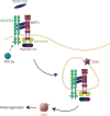
Intracellular trafficking of VEGFR2. VEGFR2 endocytosis and Neuropilin-1/synectin/myosin-VI-dependent trafficking. VEGF-A binding to VEGFR2 and Nrp1 creates a multiprotein complex and induces its endocytosis. A Nrp1-dependent trafficking that proceeds in a synectin/myosin-VI-dependent manner leads to VEGR2-driven activation of ERK signaling in EEA1+ endosomes.
Figure 2
Figure 2
Arteriogenic defects associated with delayed intracellular VEGFR2 trafficking. A. Micro-CT images of mouse coronary arteries from wild-type (top) and Synectin null (bottom mice. B. Micro-CT of mouse renal arterial circulation from wild type (top) and myosin-VI null (bottom) mice. C. Micro-CT images of mouse heart (top), hindlimb (middle) and renal (bottom) arterial circulations from mice carrying a deletion of the Nrp1 cytoplasmic domain. Adapted from Lanahan et al Dev Cell 2010 and Lanahan et al Dev Cell 2013.
Figure 3
Figure 3
Inhibition-dependent regulation of VEGFR2-driven ERK activation A. A schematic of Raf1 phosphorylation control. Dephosphorylation of Ser259 allows phosphorylation of Ser338 and activation of Raf1 kinase activity. B. Raf1 regulation of ERK, AKT and MST2 pathways cross-talk. VEGFR2-induced activation of Akt leads to MST2 phosphorylation and promotes formation of Raf1-MST2 complex that maintains Raf1 in an inactive (Ser259-phosphorylated) state. Dephosphorylation of Raf1S259 site shifts MST2 to the RASSF1A complex thereby activating LATS1 that subsequently acts on YAP. At the same time this allows phosphorylation of Raf1Ser338 thereby activating MEK/ERK signaling. Modified, in part, from. C. Increase dorsal aorta diameter in Raf1S29A (bottom) compared to WT (top) mice. D. Extra-embryonic vasculature in control (wild type) and Raf1SA259 mouse embryos. Note a marked increase in arterial size. Cx40-Connexin-40. Panels C and D adapted from
Figure 4
Figure 4. Regulation of arteriogenesis
Inflammatory (e.g. TNFα) and mechanical (e.g. shear stress) stimuli initiate arteriogenic signaling in a resting endothelial cell (top, blue). Activation of NFκB signaling by these stimuli leads to increase HIF1α and HIF2α levels, expression of adhesion receptors and production of PDGF-BB, Ang1 and Ang2. Ang2 in turn induces accumulation of a specific macrophage population that, under control of Ang1, reduce their PHD2 levels thereby increasing VEGF production. The macrophage-produced VEGF (and to a lesser extent endothelial-derived VEGF) activate arteriogenic signaling via VEGFR2/Nrp1 complex. HIF2α-induced expression of Dll4 activates Notch signaling in neighboring endothelial cells thereby controlling branching extent. PDGF-BB plays an important role in recruitment of mural cells and maturation of the new arterial network.
Figure 5
Figure 5. Key arteriogenic events
Key arteriogenic events including activation, regulation of signal transduction and arteriogenesis extent. See text for details.

References

    1. Faber JE, Chilian WM, Deindl E, van Royen N, Simons M. A brief etymology of the collateral circulation. Arterioscler Thromb Vasc Biol. 2014;34:1854–1859. - PMC - PubMed
    1. Morini MF, Dejana E. Transcriptional regulation of arterial differentiation via wnt, sox and notch. Current opinion in hematology. 2014 - PubMed
    1. Marcelo KL, Goldie LC, Hirschi KK. Regulation of endothelial cell differentiation and specification. Circulation research. 2013;112:1272–1287. - PMC - PubMed
    1. Schaper W. Collateral circulation: Past and present. Basic research in cardiology. 2009;104:5–21. - PMC - PubMed
    1. Zheng X, Xu C, Smith AO, Stratman AN, Zou Z, Kleaveland B, Yuan L, Didiku C, Sen A, Liu X, Skuli N, Zaslavsky A, Chen M, Cheng L, Davis GE, Kahn ML. Dynamic regulation of the cerebral cavernous malformation pathway controls vascular stability and growth. Developmental cell. 2012;23:342–355. - PMC - PubMed

Publication types

MeSH terms

Substances