Review
. 2015 Apr 21:9:47.
doi: 10.3389/fnana.2015.00047. eCollection 2015.

Role of developmental factors in hypothalamic function

Affiliations
Review

Role of developmental factors in hypothalamic function

Jakob Biran et al. Front Neuroanat. .

Abstract

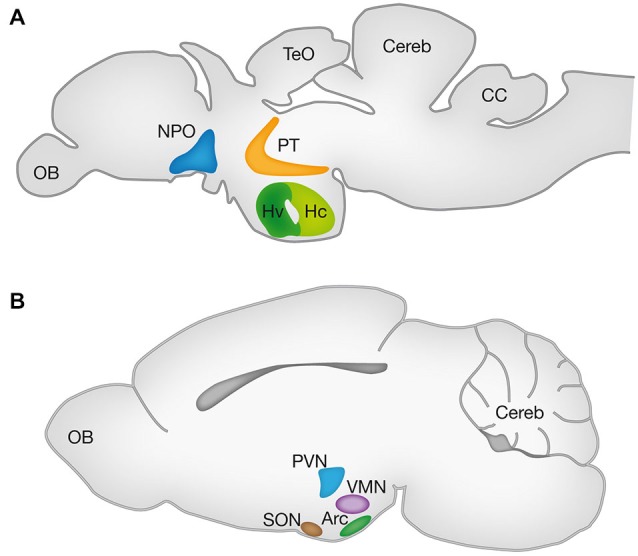
The hypothalamus is a brain region which regulates homeostasis by mediating endocrine, autonomic and behavioral functions. It is comprised of several nuclei containing distinct neuronal populations producing neuropeptides and neurotransmitters that regulate fundamental body functions including temperature and metabolic rate, thirst and hunger, sexual behavior and reproduction, circadian rhythm, and emotional responses. The identity, number and connectivity of these neuronal populations are established during the organism's development and are of crucial importance for normal hypothalamic function. Studies have suggested that developmental abnormalities in specific hypothalamic circuits can lead to obesity, sleep disorders, anxiety, depression and autism. At the molecular level, the development of the hypothalamus is regulated by transcription factors (TF), secreted growth factors, neuropeptides and their receptors. Recent studies in zebrafish and mouse have demonstrated that some of these molecules maintain their expression in the adult brain and subsequently play a role in the physiological functions that are regulated by hypothalamic neurons. Here, we summarize the involvement of some of the key developmental factors in hypothalamic development and function by focusing on the mouse and zebrafish genetic model organisms.

Keywords: Otp; PAC1; SF-1; SIM1; homeostasis; neuroendocrine; neuropeptides; zebrafish model system.


Figures

Figure 1
Figure 1
Hypothalamic nuclei in vertebrates. Schematic lateral view of the zebrafish (A) and mouse (B) brains representing the projected 2D anatomy of multiple sagittal planes. Color matched areas represents the presumed homology between specific hypothalamic areas of zebrafish and mouse (see text). Arc, arcuate nucleus; CC, crista cerebellaris; CCe, corpus cerebelli; Hv: ventral zone of periventricular hypothalamus; Hc, caudal zone of periventricular hypothalamus; NPO, neurosecretory preoptic area; OB, olfactory bulb; PT, posterior tuberculum; PVN, paraventricular nucleus; SON, supraoptic nucleus; TeO, tectum opticum; VMN, ventromedial nucleus.
Figure 2
Figure 2
Revised prosomere subdivision of the zebrafish forebrain. (A) The previously suggested zebrafish prosomeric model (see text). (B) The newly suggested zebrafish prosomeric model based on Otp and Sim1 expression patterns. In this model, the ventral boundary between the hypothalamus and prosomere 3 is shifted to the caudal limit of Sim1 and Otp domains, while the ventral posterior tuberculum (vPT), is included in the zebrafish hypothalamus. DT, dorsal thalamus; dPT, dorsal part of the posterior tuberculum; Hyp, hypothalamus; P1, prosomere 1; P2, prosomere 2; P3, prosomere 3; Pr, pretectum; Tel, telencephalon; TH, Tyrosine hydroxylase positive neurons; vPT, ventral part of the posterior tuberculum; VT, ventral thalamus.
Figure 3
Figure 3
Otp expression is maintained in the adult brain. Immunofluorescence staining of Otp (red) and oxytocin (OXT) EGFP reporter (Blechman et al., 2011) (green) in a two year-old zebrafish brain. The image shows tiled maximum intensity projection of a mid-sagital section (150 µm). Insets display separate single channel images of Otp and OXT in the NPO. CC, crista cerebellaris; CCe, corpus cerebelli; NPO, neurosecretory preoptic area; OB, olfactory bulb; Tel, telencephalon; TeO, tectum opticum. Scale bar, 200 µm.

References

    1. Acampora D., Postiglione M. P., Avantaggiato V., Di Bonito M., Vaccarino F. M., Michaud J., et al. . (1999). Progressive impairment of developing neuroendocrine cell lineages in the hypothalamus of mice lacking the Orthopedia gene. Genes Dev. 13, 2787–2800. 10.1101/gad.13.21.2787 - DOI - PMC - PubMed
    1. Achermann J. C., Ito M., Ito M., Hindmarsh P. C., Jameson J. L. (1999). A mutation in the gene encoding steroidogenic factor-1 causes XY sex reversal and adrenal failure in humans. Nat. Genet. 22, 125–126. 10.1038/9629 - DOI - PubMed
    1. Allen A. K., Spradling A. C. (2008). The Sf1-related nuclear hormone receptor Hr39 regulates Drosophila female reproductive tract development and function. Development 135, 311–321. 10.1242/dev.015156 - DOI - PubMed
    1. Altman J., Bayer S. A. (1978a). Development of the diencephalon in the rat. I. Autoradiographic study of the time of origin and settling patterns of neurons of the hypothalamus. J. Comp. Neurol. 182, 945–971. 10.1002/cne.901820511 - DOI - PubMed
    1. Altman J., Bayer S. A. (1978b). Development of the diencephalon in the rat. II. Correlation of the embryonic development of the hypothalamus with the time of origin of its neurons. J. Comp. Neurol. 182, 973–993. 10.1002/cne.901820512 - DOI - PubMed

LinkOut - more resources