

Strain US Elastography for the Characterization of Thyroid Nodules: Advantages and Limitation
- PMID: 25954310
- PMCID: PMC4411438
- DOI: 10.1155/2015/908575
Strain US Elastography for the Characterization of Thyroid Nodules: Advantages and Limitation
Abstract
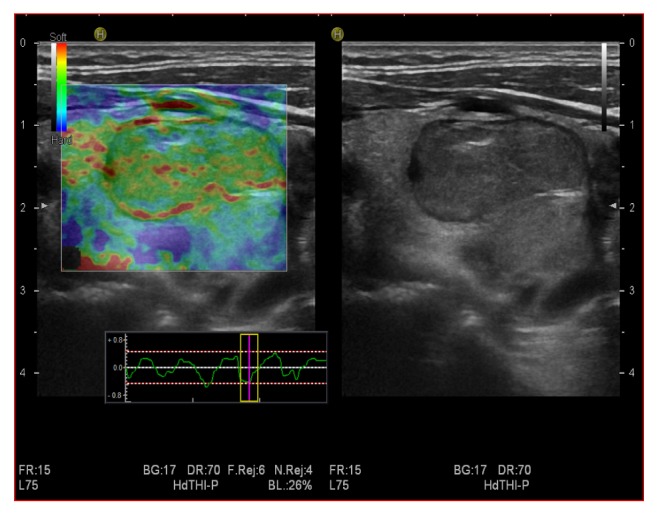
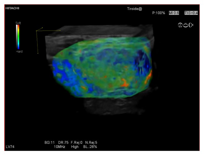
Thyroid nodules, with their high prevalence in the general population, represent a diagnostic challenge for clinicians. Ultrasound (US), although absolutely reliable in detecting thyroid nodules, is still not accurate enough to differentiate them into benign and malignant. A promising novel modality, US elastography, has been introduced in order to further increase US accuracy. The purpose of this review article is to assess the thyroid application of US strain elastography, also known as real-time elastography or quasistatic elastography. We provide a presentation of the technique, and of up-to-date literature, analyzing the most prominent results reported for thyroid nodules differentiation. The practical advantages and limitations of strain elastography are extensively discussed herein.
Figures


Similar articles
-
Ultrasound elastography in the evaluation of thyroid pathology. Current status.Eur J Radiol. 2014 Mar;83(3):420-8. doi: 10.1016/j.ejrad.2013.05.008. Epub 2013 Jun 12. Eur J Radiol. 2014. PMID: 23763859 Review.
-
US-elastography in the differential diagnosis of benign and malignant thyroid nodules.Thyroid. 2008 May;18(5):523-31. doi: 10.1089/thy.2007.0323. Thyroid. 2008. PMID: 18466077
-
Real-time ultrasound elastography for differentiation of benign and malignant thyroid nodules: a meta-analysis.J Ultrasound Med. 2014 Mar;33(3):495-502. doi: 10.7863/ultra.33.3.495. J Ultrasound Med. 2014. PMID: 24567461 Review.
-
Comparison of strain ratio with elastography score system in differentiating malignant from benign thyroid nodules.Clin Imaging. 2013 Jan-Feb;37(1):50-5. doi: 10.1016/j.clinimag.2012.04.003. Epub 2012 Jun 8. Clin Imaging. 2013. PMID: 23206607
-
Role of ultrasound elastography in prediction of malignancy in thyroid nodules.Endocr Res. 2012;37(2):67-77. doi: 10.3109/07435800.2011.633952. Endocr Res. 2012. PMID: 22489920
Cited by
-
Shear Wave Elastography versus Strain Elastography in Diagnosing Parathyroid Adenomas.Int J Endocrinol. 2020 Mar 17;2020:3801902. doi: 10.1155/2020/3801902. eCollection 2020. Int J Endocrinol. 2020. PMID: 32256571 Free PMC article.
-
Assessment of thyroid gland elasticity with shear-wave elastography in Hashimoto's thyroiditis patients.J Ultrasound. 2020 Dec;23(4):543-551. doi: 10.1007/s40477-020-00437-y. Epub 2020 Mar 17. J Ultrasound. 2020. PMID: 32185701 Free PMC article.
-
Magnetic resonance enterography (MRE) and ultrasonography (US) in the study of the small bowel in Crohn's disease: state of the art and review of the literature.Acta Biomed. 2019 Apr 24;90(5-S):38-50. doi: 10.23750/abm.v90i5-S.8337. Acta Biomed. 2019. PMID: 31085972 Free PMC article. Review.
-
Image-guided thermal ablation of benign thyroid nodules.J Ultrasound. 2016 Oct 21;20(1):11-22. doi: 10.1007/s40477-016-0221-6. eCollection 2017 Mar. J Ultrasound. 2016. PMID: 28298940 Free PMC article. Review.
-
ARFI elastography of the omentum: feasibility and diagnostic performance in differentiating benign from malignant omental masses.BMJ Open Gastroenterol. 2022 May;9(1):e000901. doi: 10.1136/bmjgast-2022-000901. BMJ Open Gastroenterol. 2022. PMID: 35523459 Free PMC article.
References
-
- Tumbridge W. M., Evered D. C., Hall R., et al. The spectrum of thyroid disease in a community: the Whick-ham survey. Clinical Endocrinology. 1997;7:481–493. - PubMed
-
- Iannuccilli J. D., Cronan J. J., Monchik J. M. Risk for malignancy of thyroid nodules as assessed by sonographic criteria: the need for biopsy. Journal of Ultrasound in Medicine. 2004;23(11):1455–1464. - PubMed
Publication types
LinkOut - more resources
Full Text Sources
Other Literature Sources
