. 2015 Apr 28:5:13.
doi: 10.1186/s13395-015-0040-z. eCollection 2015.

In vitro stability of therapeutically relevant, internally truncated dystrophins

Affiliations

In vitro stability of therapeutically relevant, internally truncated dystrophins

Jackie L McCourt et al. Skelet Muscle. .

Abstract

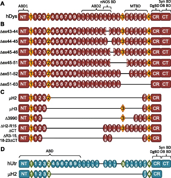
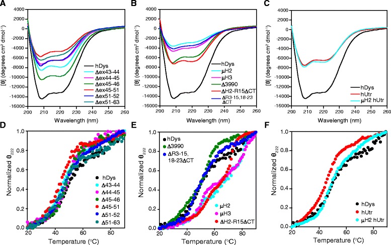
Background: The X-linked recessive disease Duchenne muscular dystrophy (DMD) is caused by mutations in the gene encoding the protein dystrophin. Despite its large size, dystrophin is a highly stable protein, demonstrating cooperative unfolding during thermal denaturation as monitored by circular dichroism spectroscopy. In contrast, internal sequence deletions have been associated with a loss of the cooperative unfolding and cause in vitro protein aggregation. Several emerging therapy options for DMD utilize internally deleted micro-dystrophins and multi-exon-skipped dystrophins that produce partially functional proteins, but the stability of such internally truncated proteins has not been investigated.

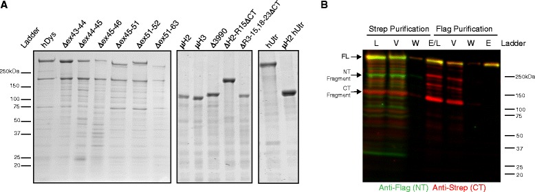
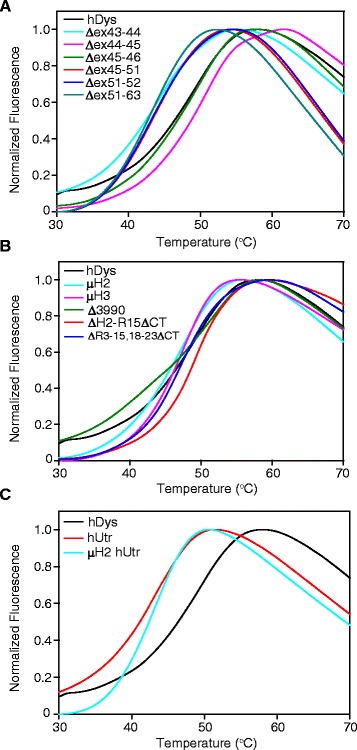
Methods: In this study, we analyzed the in vitro stability of human dystrophin constructs skipped around exon 45 or exon 51, several dystrophin gene therapy constructs, as well as human full-length and micro-utrophin. Constructs were expressed in insect cells using the baculovirus system, purified by affinity chromatography, and analyzed by high-speed sedimentation, circular dichroism spectroscopy, and differential scanning fluorimetry.

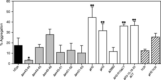
Results: Our results reveal that not all gene therapy constructs display stabilities consistent with full-length human dystrophin. However, all dystrophins skipped in-frame around exon 45 or exon 51 show stability profiles congruent with intact human dystrophin. Similar to previous studies of mouse proteins, full-length human utrophin also displays stability similar to human dystrophin and does not appear to be affected by a large internal deletion.

Conclusions: Our results suggest that the in vitro stability of human dystrophin is less sensitive to smaller deletions at natural exon boundaries than larger, more complex deletions present in some gene therapy constructs.

Keywords: Becker muscular dystrophy; Duchenne muscular dystrophy; Dystrophin; Exon skipping; Gene therapy; Utrophin.


Figures

Figure 1
Figure 1
Dystrophin and utrophin constructs analyzed. (A) Diagram of full-length human dystrophin (hDys); NT - N-terminus, CR - cysteine-rich domain, CT - C-terminus, circles - spectrin-like repeats, diamonds - unstructured ‘hinge’ regions, ABD1/2 - actin binding domains, nNOS BD - neuronal nitric oxide synthase binding domain, MTBD - microtubule binding domain, DgBD - dystroglycan binding domain, Syn BD - syntrophin binding domain, DB BD - dystrobrevin binding domain. (B) Diagrams of exon-skipped human dystrophin constructs analyzed. (C) Diagrams of gene therapy human dystrophins analyzed. (D) Diagrams of full-length human utrophin (hUtr) and a micro-utrophin (μH2 hUtr) analyzed.
Figure 2
Figure 2
Gel analysis of purified recombinant proteins. (A) Representative Coomassie-stained gels with 5 μg of exon-skipped dystrophins, gene therapy dystrophins, and utrophins loaded for comparison. (B) Western blot of purification fractions from tandem purification of dual-tagged full-length human dystrophin with N-terminal (NT) FLAG-tag (green channel) and C-terminal (CT) Strep-tag (red channel); fractions from Strep affinity purification and FLAG affinity purifications: load (L), void (V), wash (W), and elute (E). hDys - human dystrophin, hUtr - human utrophin.
Figure 3
Figure 3
Analysis of protein aggregation by high-speed sedimentation. Quantification of high-speed sedimentation assay of supernatant (S) and pellet (P) fractions where % aggregation = S/(S + P); full-length human dystrophin (hDys) in black bar, exon-skipped dystrophins in gray bars, gene therapy dystrophins in white bars, and utrophins in lined bars; *P < 0.05, **P < 0.0001 using ANOVA statistical analysis compared to full-length human dystrophin. hUtr - human utrophin.
Figure 4
Figure 4
Spectra and melt curves obtained by circular dichroism spectroscopy. (A-C) Circular dichroism absorption spectra from 200 to 260 nm for exon-skipped dystrophins (A), gene therapy constructs (B), and utrophins (C). Molar ellipticity [θ], with units of degrees centimeter squared per decimole, was calculated as θ / (10 × c × l) where c is the molar concentration of the sample (mole/L) and l is the path length in cm. (D-F) CD absorption spectra monitored at 222 nm from 20°C to 90°C for exon-skipped dystrophins (D), gene therapy dystrophins (E), and utrophins (F). Melt curves were normalized to θ222 from 0 to 1 fraction unfolded and a representative curve plotted. See Table 1 for melting temperatures (CD Tm1 and Tm2). hDys - human dystrophin, hUtr - human utrophin.
Figure 5
Figure 5
Melt curves obtained by differential scanning fluorimetry. Differential scanning fluorimetry (DSF) melt curves for exon-skipped dystrophins (A), gene therapy dystrophins (B), and utrophins (C). Fluorescence was monitored at 610 nm from 20°C to 90°C, and normalized from 0 to 1 fraction unfolded and a representative curve plotted. See Table 1 for melting temperatures (DSF Tm). hDys - human dystrophin, hUtr - human utrophin.

Similar articles

Cited by

References

    1. Hoffman EP, Brown RH, Kunkel LM. Dystrophin: the protein product of the Duchenne muscular dystrophy locus. Cell. 1987;51:919–28. doi: 10.1016/0092-8674(87)90579-4. - DOI - PubMed
    1. Muntoni F, Torelli S, Ferlini A. Review dystrophin and mutations: one gene, several proteins, multiple phenotypes. Lancet Neurol. 2003;44:731–40. doi: 10.1016/S1474-4422(03)00585-4. - DOI - PubMed
    1. Koenig M, Beggs AH, Moyer M, Scherpf S, Heindrich K, Bettecken T, et al. The molecular basis for Duchenne versus Becker muscular dystrophy: correlation of severity with type of deletion. Am J Hum Genet. 1989;45:498–506. - PMC - PubMed
    1. Rybakova IN, Ervasti JM. Dystrophin-glycoprotein complex is monomeric and stabilizes actin filaments in vitro through a lateral association. J Biol Chem. 1997;272:28771–8. doi: 10.1074/jbc.272.45.28771. - DOI - PubMed
    1. Ervasti JM. Dystrophin, its interactions with other proteins, and implications for muscular dystrophy. Biochim Biophys Acta. 2007;1772:108–17 - PubMed