

Increased NY-ESO-1 expression and reduced infiltrating CD3+ T cells in cutaneous melanoma
- PMID: 25954764
- PMCID: PMC4411457
- DOI: 10.1155/2015/761378
Increased NY-ESO-1 expression and reduced infiltrating CD3+ T cells in cutaneous melanoma
Abstract
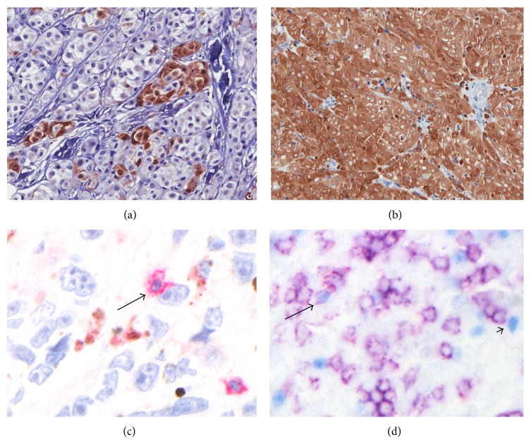
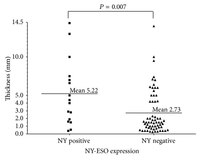
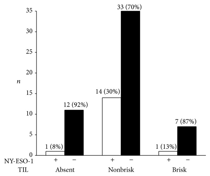
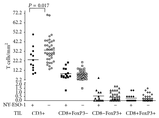
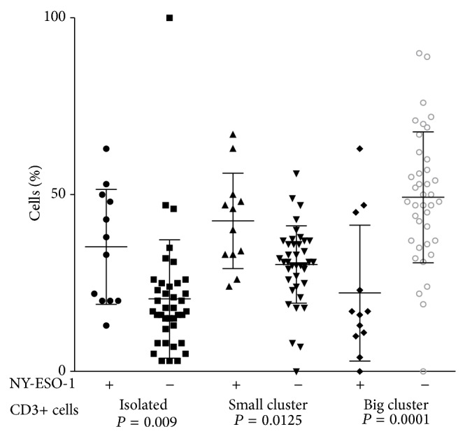
NY-ESO-1 is a cancer-testis antigen aberrantly expressed in melanomas, which may serve as a robust and specific target in immunotherapy. NY-ESO-1 antigen expression, tumor features, and the immune profile of tumor infiltrating lymphocytes were assessed in primary cutaneous melanoma. NY-ESO-1 protein was detected in 20% of invasive melanomas (16/79), rarely in in situ melanoma (1/10) and not in benign nevi (0/20). Marked intratumoral heterogeneity of NY-ESO-1 protein expression was observed. NY-ESO-1 expression was associated with increased primary tumor thickness (P = 0.007) and inversely correlated with superficial spreading melanoma (P < 0.02). NY-ESO-1 expression was also associated with reduced numbers and density of CD3+ tumor infiltrating lymphocytes (P = 0.017). When NY-ESO-1 protein was expressed, CD3+ T cells were less diffusely infiltrating the tumor and were more often arranged in small clusters (P = 0.010) or as isolated cells (P = 0.002) than in large clusters of more than five lymphocytes. No correlation of NY-ESO-1 expression with gender, age, tumor site, ulceration, lymph node sentinel status, or survival was observed. NY-ESO-1 expression in melanoma was associated with tumor progression, including increased tumor thickness, and with reduced tumor infiltrating lymphocytes.
Figures
References
-
- Armstrong B. K., Kricker A. Cutaneous melanoma. Cancer Surveys. 1994;19-20:219–240. - PubMed
-
- Pellacani G., Lo Scocco G., Vinceti M., et al. Melanoma epidemic across the millennium: time trends of cutaneous melanoma in Emilia-Romagna (Italy) from 1997 to 2004. Journal of the European Academy of Dermatology and Venereology. 2008;22(2):213–218. doi: 10.1111/j.1468-3083.2007.02388.x. - DOI - PubMed
-
- Stratigos A. J., Forsea A. M., van der Leest R. J. T., et al. Euromelanoma: a dermatology-led European campaign against nonmelanoma skin cancer and cutaneous melanoma. Past, present and future. British Journal of Dermatology. 2012;167(supplement 2):99–104. doi: 10.1111/j.1365-2133.2012.11092.x. - DOI - PubMed
Publication types
MeSH terms
Substances
LinkOut - more resources
Full Text Sources
Other Literature Sources
Medical
