. 2015 May 5;108(9):2097-102.
doi: 10.1016/j.bpj.2015.03.051.

Multidomain Assembler (MDA) Generates Models of Large Multidomain Proteins

Affiliations

Multidomain Assembler (MDA) Generates Models of Large Multidomain Proteins

Samuel Hertig et al. Biophys J. .

Abstract

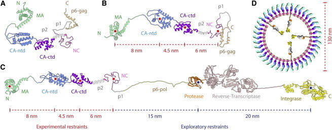
Homology modeling predicts protein structures using known structures of related proteins as templates. We developed MULTIDOMAIN ASSEMBLER (MDA) to address the special problems that arise when modeling proteins with large numbers of domains, such as fibronectin with 30 domains, as well as cases with hundreds of templates. These problems include how to spatially arrange nonoverlapping template structures, and how to get the best template coverage when some sequence regions have hundreds of available structures while other regions have a few distant homologs. MDA automates the tasks of template searching, visualization, and selection followed by multidomain model generation, and is part of the widely used molecular graphics package UCSF CHIMERA (University of California, San Francisco). We demonstrate applications and discuss MDA's benefits and limitations.


Figures

Figure 1
Figure 1
(A) Schematic structure of the 30 domains of fibronectin (Fn) with three domain types: Fn type I (FnI, circles), Fn type II (FnII, hexagons), and Fn type III (FnIII, rectangles). (Open) Domains in human Fn whose structures are not available in the PDB. (B) Schematic structure of HIV Gag and GagPol polyproteins. Gag and GagPol share the MA domain, the N-terminal and C-terminal CA domains (CAn and CAc), and the NC domain. The p6 domain of Gag differs from the p6 domain of GagPol, and GagPol has the additional domains Protease (PR), Reverse-Transcriptase (RT), and Integrase (Int). Structures of fragments of entire domains have been determined for several HIV strains. (C) Schematic overview of the MDA pipeline (dotted circles indicate selections made by MDA or the user at each step; see main text for details). To see this figure in color, go online.
Figure 2
Figure 2
(A) Structural overview of Fn domain structures (templates) arranged from N to C with homologs stacked downward by filtered ranking. (Gray) Residues that aligned in BLAST; (black) residues that did not align; (red) residues that differ from the target sequence; and (blue) co-complexed chains. (Gray spheres) Gaps with no structural coverage (volumes proportional to missing residues). Image saved from CHIMERA with ambient-only lighting and silhouettes. (B) Eliminating all but the structures with highest percent identity per residue, allowing multiple hits per PDB, and manually removing a few redundant templates results in a different ensemble with 14 templates and smaller gaps. (C) A model of monomeric Fn produced by MDA using MODELLER and the templates shown in (B) (see Table S1 and Fig. S1 for a detailed protocol) can be compared with experimental data or serve as an input structure for subsequent computational modeling approaches. The overall length of the model measures 90 nm, and the distance between FnIII-7 and FnIII-15 measures 29 nm, which agrees with literature values (25). (D) The same model shown in surface representation and colored by electrostatic potential (red, −10 kcal/mole; white, 0; blue, +10 kcal/mole). To see this figure in color, go online.
Figure 3
Figure 3
MDA output structures of Gag polyproteins with (A) no restraints, and (B) three distance restraints derived from electron microscopy data (26) (indicated with red dotted lines) applied at the geometric centers of domains (red dots). (C) An MDA output of GagPol with two exploratory restraints (shown as blue dotted lines) added, generates an elongated molecule while respecting bond lengths. The model visually reveals regions of interest such as the huge linker (56 amino acids), consisting of a structurally plausible trans-frame peptide, and p6-pol (believed to be disordered), connecting Gag and the protease and regions of concern such as the structurally unlikely straight sequence connecting Reverse-Transcriptase with Integrase. These restraints change the shape of the resulting model to coarsely approximate the linear arrangement found in the lattice of immature virions as visually hypothesized with the sketch in (D). To see this figure in color, go online.

Similar articles

Cited by

References

    1. Levitt M. Nature of the protein universe. Proc. Natl. Acad. Sci. USA. 2009;106:11079–11084. - PMC - PubMed
    1. Schwede T. Protein modeling: what happened to the “protein structure gap”? Structure. 2013;21:1531–1540. - PMC - PubMed
    1. Shapovalov M.V., Wang Q., Dunbrack R.L., Jr. BIOASSEMBLYMODELER (BAM): user-friendly homology modeling of protein homo- and heterooligomers. PLoS ONE. 2014;9:e98309. - PMC - PubMed
    1. Söding J., Biegert A., Lupas A.N. The HHPRED interactive server for protein homology detection and structure prediction. Nucleic Acids Res. 2005;33:W244–W248. - PMC - PubMed
    1. Roy A., Kucukural A., Zhang Y. I-TASSER: a unified platform for automated protein structure and function prediction. Nat. Protoc. 2010;5:725–738. - PMC - PubMed

Publication types