. 2015 May 5;108(9):2212-22.
doi: 10.1016/j.bpj.2015.03.056.

Ordered raft domains induced by outer leaflet sphingomyelin in cholesterol-rich asymmetric vesicles

Affiliations

Ordered raft domains induced by outer leaflet sphingomyelin in cholesterol-rich asymmetric vesicles

Qingqing Lin et al. Biophys J. .

Abstract

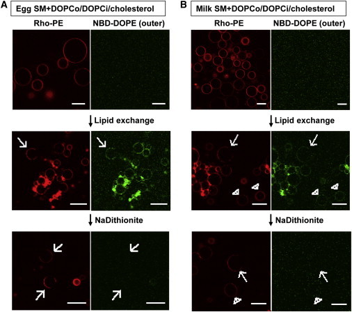
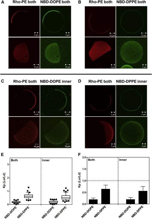
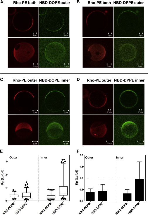
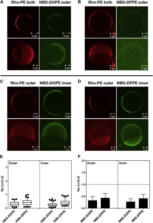
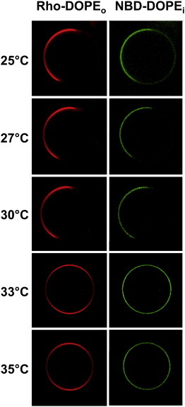
Sphingolipid- and cholesterol-rich liquid-ordered (Lo) lipid domains (rafts) are thought to be important organizing elements in eukaryotic plasma membranes. How they form in the sphingolipid-poor cytosolic (inner) membrane leaflet is unclear. Here, we characterize how outer-leaflet Lo domains induce inner-leaflet-ordered domains, i.e., interleaflet coupling. Asymmetric vesicles studied contained physiologically relevant cholesterol levels (∼ 37 mol %), a mixture of SM (sphingomyelin) and DOPC (dioleoylphosphatidylcholine) in their outer leaflets, and DOPC in their inner leaflets. Lo domains were observed in both leaflets, and were in register, indicative of coupling between SM-rich outer-leaflet-ordered domains and inner-leaflet-ordered domains. For asymmetric vesicles with outer-leaflet egg SM or milk SM, a fluorescent lipid with unsaturated acyl chains (NBD-DOPE) was depleted in both the outer- and inner-leaflet-ordered domains. This suggests the inner-leaflet-ordered domains were depleted in unsaturated lipid (i.e., DOPC) and thus rich in cholesterol. For asymmetric vesicles containing egg SM, outer-leaflet Lo domains were also depleted in a saturated fluorescent lipid (NBD-DPPE), while inner-leaflet Lo domains were not. This indicates that inner- and outer-leaflet Lo domains can have significantly different physical properties. In contrast, in asymmetric vesicles containing outer-leaflet milk SM, which has long acyl chains capable of interdigitating into the inner leaflet, both outer- and inner-leaflet Lo domains were depleted, to a similar extent, in NBD-DPPE. This is indicative of interdigitation-enhanced coupling resulting in inner- and outer-leaflet Lo domains with similar physical properties.


Figures

Figure 1
Figure 1
Preparation of SM + DOPCo/DOPCi/∼37 mol % cholesterol vesicles. Asymmetric GUVs composed 37 mol % cholesterol and an inner leaflet of DOPC and an outer leaflet of egg SM (A) or milk SM (B) and DOPC. The acceptor vesicles were labeled with 0.02 mol % Rho-DOPE and the donor vesicles were labeled with 0.01 mol % NBD-DOPE. (Top) Before exchange. (Middle) After exchange. (Bottom) After lipid exchange and 1 mg/mL sodium dithionite added to destroy outer-leaflet NBD fluorescence. (Arrows) Vesicles with large-scale domain formation, which are the type of vesicles used for the later Kp analysis; (arrow heads) some homogeneous vesicles without domain formation. Vesicle positions are altered by dithionite addition and washing, so the middle and bottom panels do not represent the same vesicles. Scale bar = 20 μm. To see this figure in color, go online.
Figure 2
Figure 2
Images and NBD partition data for symmetric vesicles composed of 1:1 (mol/mol) egg SM/DOPC with 37 mol % cholesterol. Vesicles were labeled with Rho-DOPE and NBD-DOPE (A) or NBD-DPPE (B) in both leaflets or with Rho-DOPE in both leaflets and NBD-DOPE (C) or NBD-DPPE (D) in the inner leaflet. (Outer-leaflet NBD was reduced with sodium dithionite to restrict NBD fluorescence to the inner leaflet.) Two-dimensional cross-section images (upper panels in A–D) and three-dimensional reconstruction images (bottom panels in A–D) shown. The two- and three-dimensional images are from different vesicles. (E and F) Box plot and bar graph representations, respectively, of the NBD-DOPE and NBD-DPPE partition coefficient Kp (Lo/Ld). NBD-DOPE in both leaflets, n = 21; NBD-DOPE in the inner leaflet, n = 27; NBD-DPPE in both leaflets, n = 27; and NBD-DPPE in the inner leaflet, n = 33. The value of n here and below equals the number of vesicles analyzed. Kp (Lo/Ld) is the ratio of NBD fluorescence in Lo domains divided by that in Ld domains; Kp estimated here and in the following figures used intensity at 3–5 positions within Ld regions and 3–5 positions within Lo regions in each vesicle. To see this figure in color, go online.
Figure 3
Figure 3
Images and NBD partition data for asymmetric egg SM + DOPCo/DOPCi/∼37 mol % cholesterol GUV. GUV labeled with (A) NBD-DOPE or (B) NBD-DPPE in the outer leaflet, and labeled with NBD-DOPE (C) or with NBD-DPPE (D) in the inner leaflet. (C and D) To restrict NBD fluorescence to the inner leaflet, outer leaflet NBD was reduced with sodium dithionite. For GUV with outer-leaflet NBD lipid, Rho-DOPE was in both leaflets; for GUV with inner-leaflet NBD, lipid Rho-DOPE was exchanged into the outer leaflet. Note that the vesicles in (D) are from the less abundant subpopulation in which NBD-DPPE preferentially associates with Lo domains. (A–D) Two-dimensional cross-section images (upper panel) and three-dimensional reconstruction images (bottom panel) are shown. Two- and three-dimensional images are from different vesicles. (E and F) Box plot and bar graph representations, respectively, of NBD-DOPE and NBD-DPPE Kp (Lo/Ld). Box boundaries at 25% and 75% values, whiskers set at 5% and 95% values. NBD-DOPE in the outer leaflets, n = 23; NBD-DOPE in the inner leaflet, n = 37; NBD-DPPE in the outer leaflet, n = 46; NBD-DPPE in the inner leaflet, n = 44. To see this figure in color, go online.
Figure 4
Figure 4
Images and NBD partition data for asymmetric milk SM + DOPCo/DOPCi/∼37 mol % cholesterol GUV. GUV labeled with (A) NBD-DOPE or (B) NBD-DPPE in the outer leaflet, and with NBD-DOPE (C) or NBD-DPPE (D) in the inner leaflet. (C and D) To restrict NBD fluorescence to the inner leaflet, outer leaflet NBD was reduced with sodium dithionite. For GUV with outer-leaflet NBD lipid, Rho-DOPE was in both leaflets; for GUV with inner-leaflet NBD lipid, Rho-DOPE was exchanged into their outer leaflet. (A–D) Two-dimensional cross-section images (upper panel) and three-dimensional reconstruction images (bottom panel) were shown. Two- and three-dimensional images were taken from different vesicles except for (B), in which they are from the same vesicle to allow visualization of NBD partitioning. (E and F) Box plot and bar graph representations, respectively, of NBD-DOPE and NBD-DPPE Kp (Lo/Ld). NBD-DOPE in the outer leaflets, n = 18; NBD-DOPE in the inner leaflet, n = 22; NBD-DPPE in the outer leaflet, n = 17; NBD-DPPE in the inner leaflet, n = 18. To see this figure in color, go online.
Figure 5
Figure 5
Thermal stability of large domains in egg SM + DOPCo/DOPCi/∼37 mol % cholesterol GUVs. Acceptor vesicles contained 63:37 (mol/mol) DOPC/cholesterol labeled with NBD-DOPE. Donor vesicles contained 63:37 (mol/mol) egg SM/cholesterol labeled with Rho-DOPE. NBD in the outer leaflet was reduced with sodium dithionite for 90 s to restrict NBD fluorescence to the inner leaflet. Images were taken by the LAS AF confocal microscope (Leica Microsystems) with the CUBE & BOX temperature control system. Sample temperature was increased in steps taking a few minutes, with micrographs recorded 1–2 min after the desired temperature was reached. To see this figure in color, go online.

Comment in

  • Of rafts and lipid chain lengths.
    Hristova K, Hinderliter A. Hristova K, et al. Biophys J. 2015 May 5;108(9):2096. doi: 10.1016/j.bpj.2015.03.057. Biophys J. 2015. PMID: 25954867 Free PMC article. No abstract available.

Similar articles

Cited by

References

    1. Bretscher M.S. Asymmetrical lipid bilayer structure for biological membranes. Nat. New Biol. 1972;236:11–12. - PubMed
    1. Devaux P.F. Static and dynamic lipid asymmetry in cell membranes. Biochemistry. 1991;30:1163–1173. - PubMed
    1. Brown D.A., London E. Structure and origin of ordered lipid domains in biological membranes. J. Membr. Biol. 1998;164:103–114. - PubMed
    1. van Meer G., Voelker D.R., Feigenson G.W. Membrane lipids: where they are and how they behave. Nat. Rev. Mol. Cell Biol. 2008;9:112–124. - PMC - PubMed
    1. Collins M.D. Interleaflet coupling mechanisms in bilayers of lipids and cholesterol. Biophys. J. 2008;94:L32–L34. - PMC - PubMed

Publication types

MeSH terms