. 2015 May 8;10(5):e0126371.
doi: 10.1371/journal.pone.0126371. eCollection 2015.

Canine Mammary Tumours Are Affected by Frequent Copy Number Aberrations, including Amplification of MYC and Loss of PTEN

Affiliations

Canine Mammary Tumours Are Affected by Frequent Copy Number Aberrations, including Amplification of MYC and Loss of PTEN

Kaja S Borge et al. PLoS One. .

Abstract

Background: Copy number aberrations frequently occur during the development of many cancers. Such events affect dosage of involved genes and may cause further genomic instability and progression of cancer. In this survey, canine SNP microarrays were used to study 117 canine mammary tumours from 69 dogs.

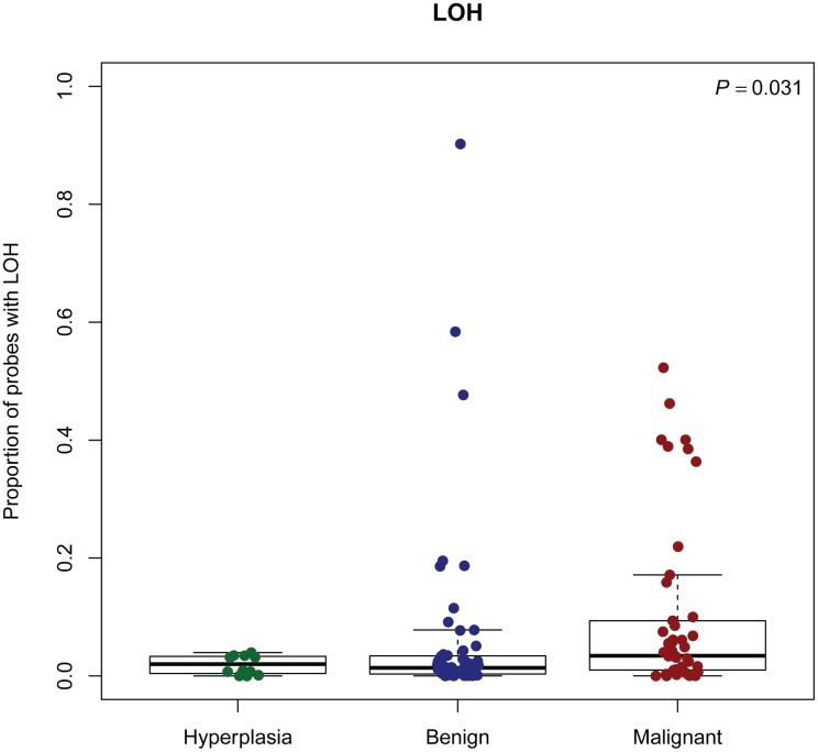
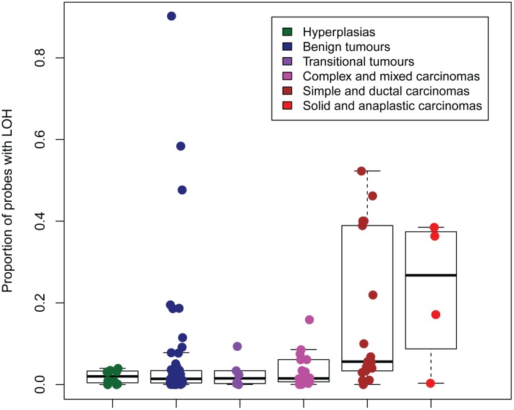
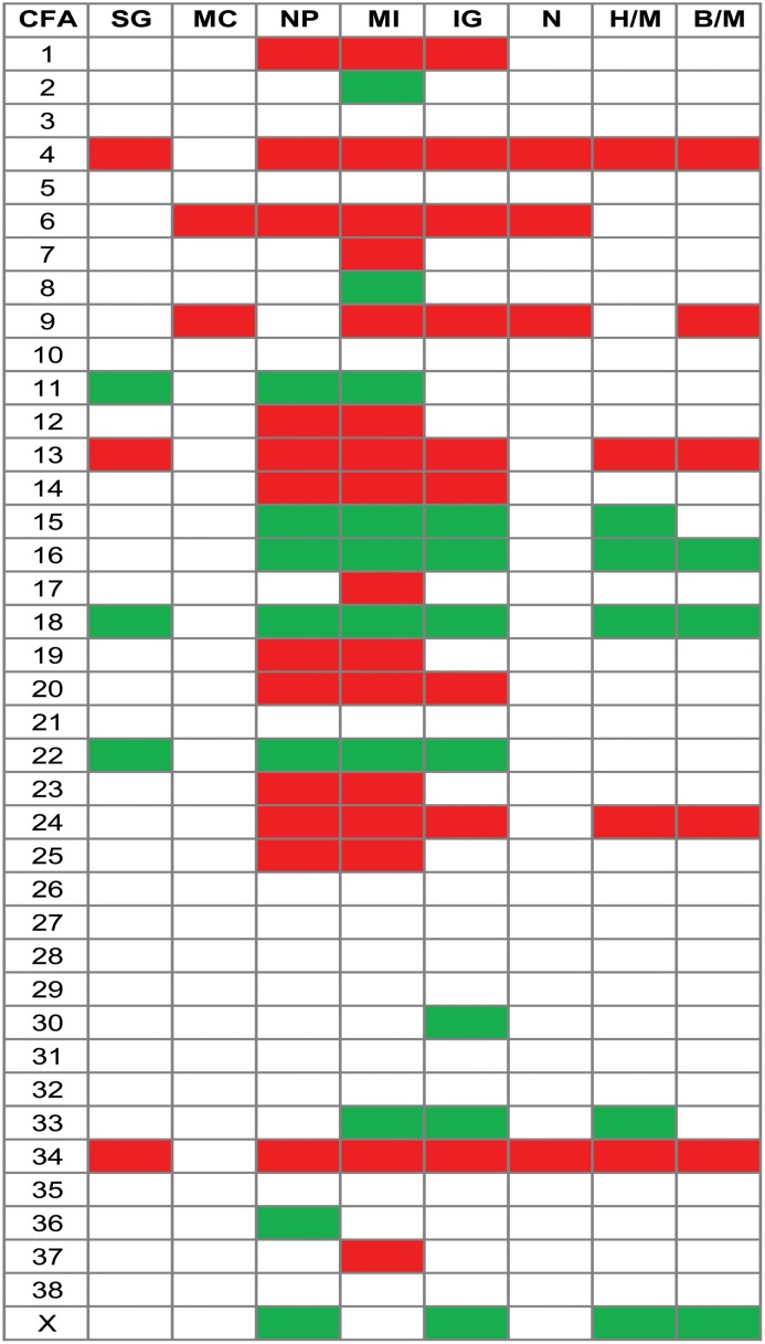
Results: We found a high occurrence of copy number aberrations in canine mammary tumours, losses being more frequent than gains. Increased frequency of aberrations and loss of heterozygosity were positively correlated with increased malignancy in terms of histopathological diagnosis. One of the most highly recurrently amplified regions harbored the MYC gene. PTEN was located to a frequently lost region and also homozygously deleted in five tumours. Thus, deregulation of these genes due to copy number aberrations appears to be an important event in canine mammary tumour development. Other potential contributors to canine mammary tumour pathogenesis are COL9A3, INPP5A, CYP2E1 and RB1. The present study also shows that a more detailed analysis of chromosomal aberrations associated with histopathological parameters may aid in identifying specific genes associated with canine mammary tumour progression.

Conclusions: The high frequency of copy number aberrations is a prominent feature of canine mammary tumours as seen in other canine and human cancers. Our findings share several features with corresponding studies in human breast tumours and strengthen the dog as a suitable model organism for this disease.


Conflict of interest statement

Competing Interests: The authors have declared that no competing interests exist.

Figures

Fig 1
Fig 1. ASCAT estimate of ploidy in different subgroups of tumours.
Hyperplasias (n = 12), benign (n = 55) and malignant tumours (n = 46). For the graphical presentation, the ploidy estimates were rounded to the nearest whole number.
Fig 2
Fig 2. ASCAT estimate of percentage of aberrant cells in different subtypes of tumours.
Hyperplasias (n = 12), benign tumours (n = 55) and malignant tumours (n = 46).
Fig 3
Fig 3. Recurring gains and losses across all tumour samples, according to ASCAT analysis.
The figure shows recurring gains and losses across all tumour samples, relative to the ASCAT-estimate of ploidy for each tumour. Red = gains, green = losses, chromosome number along the x-axis, and frequency of tumours with aberration on the y-axis. Peaks outside the dotted blue lines are aberrations found in 20 or more samples.
Fig 4
Fig 4. Recurring gains and losses in three subgroups of CMTs, according to ASCAT analysis.
The figure shows recurring gains and losses in three subgroups of CMTs, relative to the ASCAT-estimate of ploidy for each tumour. Red = gains, green = losses, chromosome number along the x-axis, and frequency of tumours with aberration on the y-axis.
Fig 5
Fig 5. Proportion of probes with LOH in subgroups of CMTs, according to ASCAT analysis.
LOH: Loss of heterozygosity. P: P-value from Kruskal-Wallis test.
Fig 6
Fig 6. Proportion of probes with LOH in groups of morphologically similar tumours, according to ASCAT analysis.
LOH: Loss of heterozygosity.
Fig 7
Fig 7. Chromosome-wise gains (red) and losses (green) for different histopathological parameters.
Gain/loss frequencies from the ASCAT analysis were used for these calculations. Only chromosomes with regions with a CNA-frequency difference ≥20% are shown (see Materials and Methods). The corresponding contrasts between hyperplasias/malignant tumours and benign/malignant tumours are shown for comparison. There were no regions with difference in frequency of loss ≥20% for compared categories of the parameters myoepithelial cells and necrosis. CFA: canine chromosome. SG: Solid growth (no/yes), MC: Myoepithelial cells (yes/no), NP: Nuclear pleomorphism (no/moderate-severe), MI: Mitotic index (0/above 9 pr HBF), IG: Invasive growth, tumour stroma (no/yes), N: Necrosis (no/yes), H/M: Hyperplasia vs malignant, B/M: Benign vs malignant.

Similar articles

Cited by

References

    1. Shearin AL, Ostrander EA. Leading the way: canine models of genomics and disease. Dis Model Mech. 2010; 3: 27–34. 3/1-2/27 [pii];10.1242/dmm.004358 - DOI - PMC - PubMed
    1. Bronson RT. Variation in age at death of dogs of different sexes and breeds. Am J Vet Res. 1982; 43: 2057–2059. - PubMed
    1. Bronden LB, Nielsen SS, Toft N, Kristensen AT. Data from the Danish veterinary cancer registry on the occurrence and distribution of neoplasms in dogs in Denmark. Vet Rec. 2010; 166: 586–590. 10.1136/vr.b4808 - DOI - PubMed
    1. Boldizsar H, Szenci O, Muray T, Csenki J. Studies on canine mammary tumours. I. Age, seasonal and breed distribution. Acta Vet Hung. 1992; 40: 75–87. - PubMed
    1. Egenvall A, Bonnett BN, Ohagen P, Olson P, Hedhammar A, von Euler H. Incidence of and survival after mammary tumors in a population of over 80,000 insured female dogs in Sweden from 1995 to 2002. Prev Vet Med. 2005; 69: 109–127. - PubMed

Publication types

MeSH terms

Substances