Randomized Controlled Trial
. 2015 May 8;10(5):e0124987.
doi: 10.1371/journal.pone.0124987. eCollection 2015.

Characterization of early disease status in treatment-naive male paediatric patients with Fabry disease enrolled in a randomized clinical trial

Affiliations
Randomized Controlled Trial

Characterization of early disease status in treatment-naive male paediatric patients with Fabry disease enrolled in a randomized clinical trial

Frits A Wijburg et al. PLoS One. .

Abstract

Trial design: This analysis characterizes the degree of early organ involvement in a cohort of oligo-symptomatic untreated young patients with Fabry disease enrolled in an ongoing randomized, open-label, parallel-group, phase 3B clinical trial.

Methods: Males aged 5-18 years with complete α-galactosidase A deficiency, without symptoms of major organ damage, were enrolled in a phase 3B trial evaluating two doses of agalsidase beta. Baseline disease characteristics of 31 eligible patients (median age 12 years) were studied, including cellular globotriaosylceramide (GL-3) accumulation in skin (n = 31) and kidney biopsy (n = 6; median age 15 years; range 13-17 years), renal function, and glycolipid levels (plasma, urine).

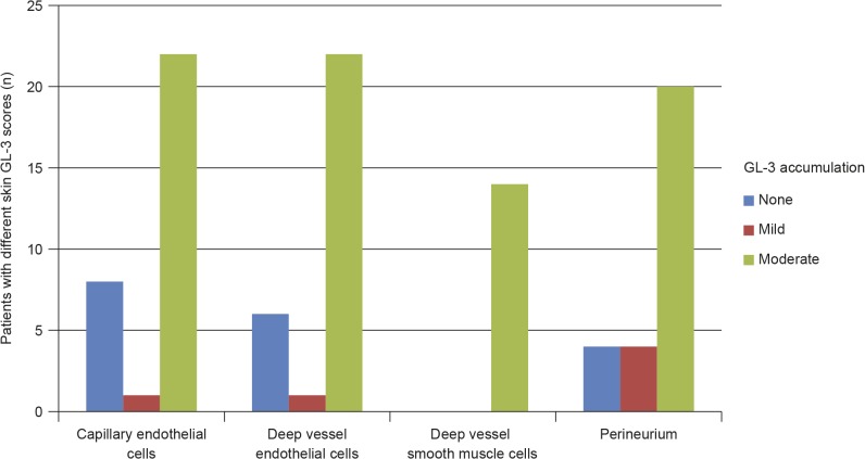
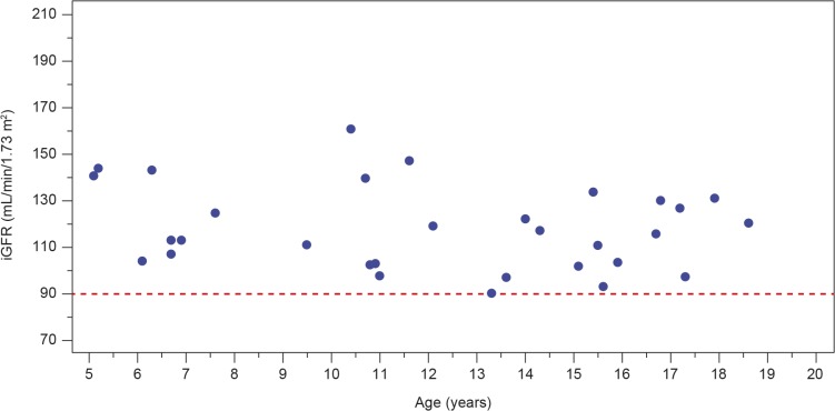
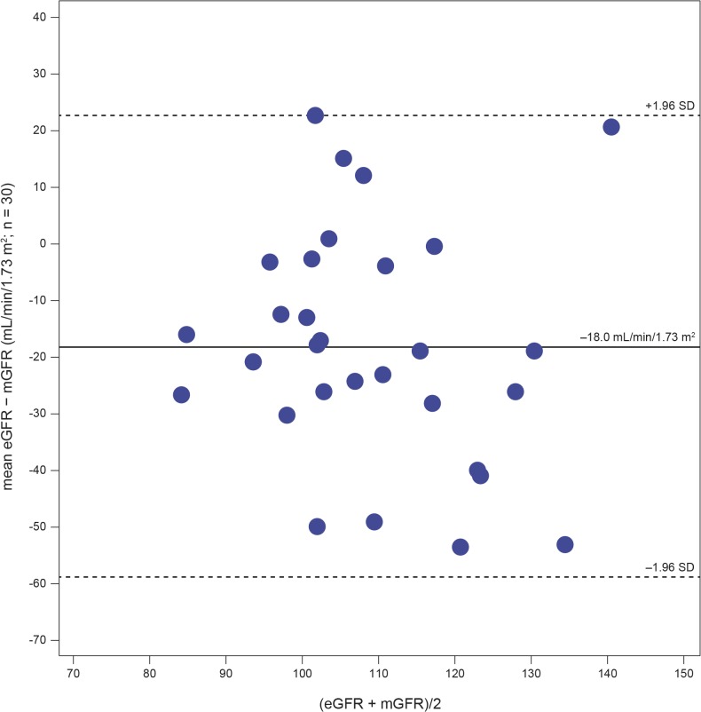
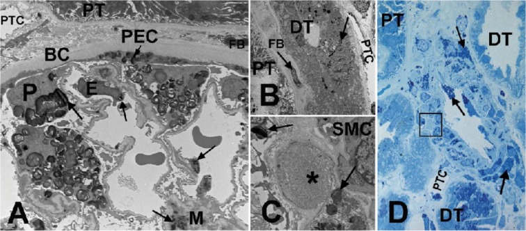
Results: Plasma and urinary GL-3 levels were abnormal in 25 of 30 and 31 of 31 patients, respectively. Plasma lyso-GL-3 was elevated in all patients. GL-3 accumulation was documented in superficial skin capillary endothelial cells (23/31 patients) and deep vessel endothelial cells (23/29 patients). The mean glomerular filtration rate (GFR), measured by plasma disappearance of iohexol, was 118.1 mL/min/1.73 m(2) (range 90.4-161.0 mL/min/1.73 m(2)) and the median urinary albumin/creatinine ratio was 10 mg/g (range 4.0-27.0 mg/g). On electron microscopy, renal biopsy revealed GL-3 accumulation in all glomerular cell types (podocytes and parietal, endothelial, and mesangial cells), as well as in peritubular capillary and non-capillary endothelial, interstitial, vascular smooth muscle, and distal tubules/collecting duct cells. Lesions indicative of early Fabry arteriopathy and segmental effacement of podocyte foot processes were found in all 6 patients.

Conclusions: These data reveal that in this small cohort of children with Fabry disease, histological evidence of GL-3 accumulation, and cellular and vascular injury are present in renal tissues at very early stages of the disease, and are noted before onset of microalbuminuria and development of clinically significant renal events (e.g. reduced GFR). These data give additional support to the consideration of early initiation of enzyme replacement therapy, potentially improving long-term outcome.

Trial registration: ClinicalTrials.gov NCT00701415.


Conflict of interest statement

Competing Interests: U.R. has read the journal's policy and the authors of this manuscript have the following competing interests: F.A.W. and D.G.B. have received honoraria for presentations and board meetings, travel expenses to meetings and honoraria for consultancy work from Genzyme and Shire HGT, and have received unrestricted educational grants and research grants from Genzyme. L.C. has received speakers’ fees and travel support from Genzyme. B.B., B.L.T., and K.A.H. are full-time employees of Genzyme, the funder of this study. C.T. has received speakers’ fees and travel support from Genzyme and Shire HGT. R.J.H. consults with Genzyme and Shire HGT and has been an investigator in clinical trials sponsored by Genzyme, Shire HGT, and Amicus. These activities have been monitored and found to be in compliance with the conflict of interest policies at Cincinnati Children’s Hospital Medical Center. G.D. has received honoraria and grants for presentations and meetings from Genzyme and Shire HGT. A.F. has received honoraria for presentations and board meetings, and has received unrestricted educational grants and research grants from Genzyme and Shire HGT. M.M. is a member of the Genzyme-sponsored FDA-mandated Fabry Registry Board, has an investigator-initiated research grant from Genzyme, is a consultant to Genzyme for clinical trial design, and has received speakers’ fees and travel support from Genzyme. These interests have been reviewed and managed by the University of Minnesota in accordance with its conflict of interest policies. A.T.S. has received honoraria for presentations and board meetings and travel expenses to meetings from Genzyme and Shire HGT. C.R.S. is a consultant to Genzyme and has received research funding and educational grants from Genzyme. S.P.S. has been a site primary investigator in clinical trials, received research support and educational grants sponsored by Genzyme, Shire, Protalix, Actelion and Amicus. S.P.S. has also received honorarium and travel support as speaker and for investigator meetings of Genzyme, Shire HGT and Protalix. These activities have been monitored and found to be in compliance with the conflict of interest policies at Emory University. B.N. has received research grants, speaker's honoraria and travel support from Genzyme, is a consultant to Genzyme and Amicus, is on medical advisory board of Amicus, and received research grant from Roche. These interests have been reviewed and managed by the University of Washington Office of Research. U.R. has received research and travel grants and honoraria for lectures from Shire HGT and Genzyme. C.F. has been an investigator in clinical trials sponsored by Genzyme. The authors received no honoraria related to the development of this publication. A.F. has no conflicts of interest. This does not alter the authors' adherence to all the PLOS ONE policies on sharing data and materials, as detailed online in the guide for authors.

Figures

Fig 1
Fig 1. Skin GL-3 scores in treatment-naive male paediatric patients with Fabry disease.
Data missing for deep vessel endothelial cells (n = 2), deep vessel smooth muscle cells (n = 17), and perineurium (n = 3); cell types not present in biopsy. GL-3 = globotriaosylceramide.
Fig 2
Fig 2. Levels of glomerular filtration rate measured by plasma disappearance of iohexol (iGFR) in treatment-naive male paediatric patients with Fabry disease.
The red dashed line indicates the iGFR inclusion criterion for the study (90 mL/min/1.73 m2). iGFR = glomerular filtration rate as measured by plasma disappearance of iohexol.
Fig 3
Fig 3. Bland–Altman plot showing the uncertainty for the eGFR Bedside Schwartz equation.
GFR = glomerular filtration rate; eGFR = GFR estimated from serum creatinine levels; mGFR = measured GFR; SD = standard deviation.
Fig 4
Fig 4. Kidney biopsy images from a male patient with Fabry disease.
(A) EM of a glomerulus. Arrows show GL-3 inclusions in podocytes (P), endothelial cells (E), mesangial cells (M), and parietal epithelial cells (PEC). (B) EM of a distal tubule (DT) with GL-3 inclusions (arrows) accumulated in its epithelial cells, in contrast to the adjacent proximal tubule (PT) with no obvious GL-3 inclusion. (C) EM close-up of the square shown in (D), displaying early Fabry arteriopathy (asterisk) focally replacing smooth muscle cells (SMC) of an arteriolar wall. Arrows show GL-3 inclusions in adjacent smooth muscle cells. (D) Methylene blue/Azure II (Richardson’s) stained semi-thin section of an arteriole. Note that the arteriopathy shown in (C) with higher magnification EM is not easily identifiable by LM (square). Arrows show GL-3 inclusions in endothelial cells and smooth muscle cells of the artery. BC = Bowman's capsule; DT = distal tubule; E = endothelial cells; EM = electron microscopy; FB = fibroblast; GL-3 = globotriaosylceramide; LM = light microscopy; M = mesangial cells; P = podocytes; PEC = parietal epithelial cells; PT = proximal tubule; PTC = peritubular capillary; SMC = smooth muscle cells. EM measurements of GL-3 deposition in glomerular cell types are shown in Table 4 [16]. Unbiased morphometric EM estimates of GL-3 in the three glomerular cell types including Vv (Inc/PC), Vv(Inc/Endo), and Vv(Inc/Mes) [16,27], as well as podocyte foot process width and percentage glomerular capillary endothelial fenestration [16,26,27] are tabulated in Table 4.

References

    1. Desnick RJ, Ioannou YA, Eng CM. Alpha-galactosidase A deficiency: Fabry Disease In: Scriver C, Beaudet A, Sly W, Valle D, editors. The metabolic bases of inherited disease. 8th ed. New York: McGraw-Hill; 2001. pp. 3733–3774.
    1. Breunig F, Wanner C. Enzyme replacement therapy for Fabry disease: proving the clinical benefit. Nephrol Dial Transplant. 2003;18: 7–9. - PubMed
    1. Eng CM, Germain DP, Banikazemi M, Warnock DG, Wanner C, Hopkin RJ, et al. Fabry disease: guidelines for the evaluation and management of multi-organ system involvement. Genet Med. 2006;8: 539–548. - PubMed
    1. Wilcox WR, Oliviera JP, Hopkin RJ, Ortiz A, Banikazemi M, Feldt-Rasmussen U, et al. Females with Fabry disease frequently have major organ involvement: lessons from the Fabry registry. Mol Genet Metab. 2008;93: 112–128. - PubMed
    1. Schiffmann R, Warnock DG, Banikazemi M, Bultas J, Linthorst GE, Packman S, et al. Fabry disease: progression of nephropathy, and prevalence of cardiac and cerebrovascular events before enzyme replacement therapy. Nephrol Dial Transplant. 2009;24: 2102–2111. 10.1093/ndt/gfp031 - DOI - PMC - PubMed

Publication types

MeSH terms

Associated data