Review
. 2015 Jul;185(7):1800-8.
doi: 10.1016/j.ajpath.2015.03.009. Epub 2015 May 5.

Multiple Mechanisms of Unfolded Protein Response-Induced Cell Death

Affiliations
Review

Multiple Mechanisms of Unfolded Protein Response-Induced Cell Death

Nobuhiko Hiramatsu et al. Am J Pathol. 2015 Jul.

Abstract

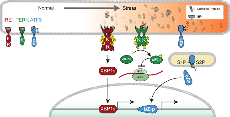
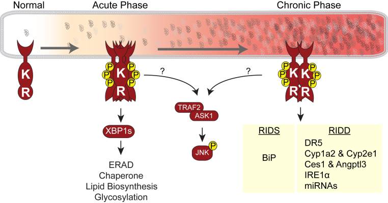
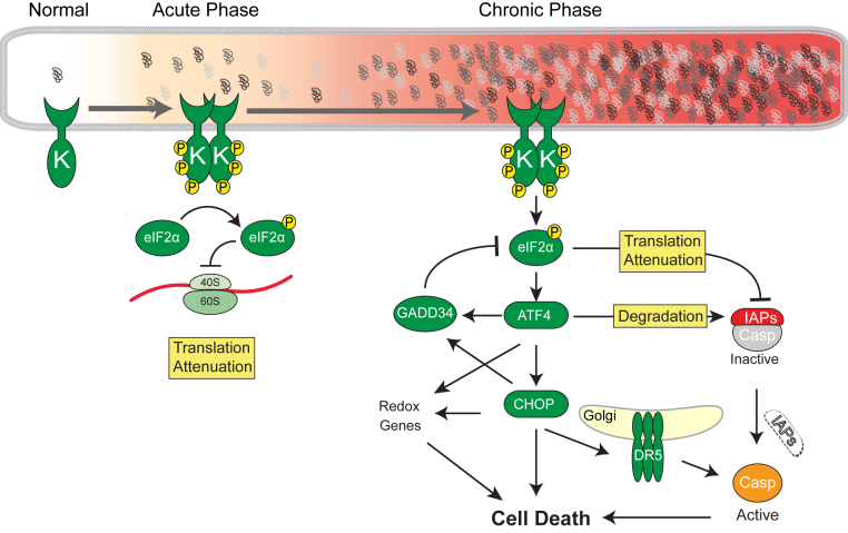
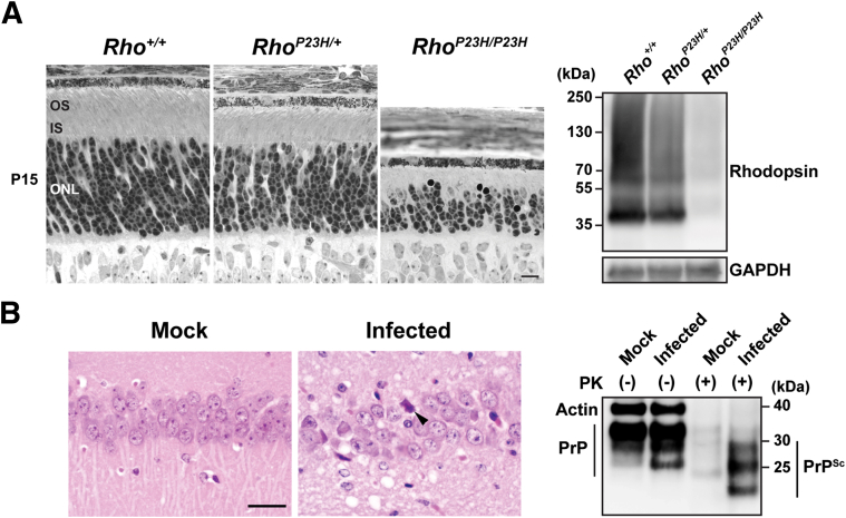
Eukaryotic cells fold and assemble membrane and secreted proteins in the endoplasmic reticulum (ER), before delivery to other cellular compartments or the extracellular environment. Correctly folded proteins are released from the ER, and poorly folded proteins are retained until they achieve stable conformations; irreparably misfolded proteins are targeted for degradation. Diverse pathological insults, such as amino acid mutations, hypoxia, or infection, can overwhelm ER protein quality control, leading to misfolded protein buildup, causing ER stress. To cope with ER stress, eukaryotic cells activate the unfolded protein response (UPR) by increasing levels of ER protein-folding enzymes and chaperones, enhancing the degradation of misfolded proteins, and reducing protein translation. In mammalian cells, three ER transmembrane proteins, inositol-requiring enzyme-1 (IRE1; official name ERN1), PKR-like ER kinase (PERK; official name EIF2AK3), and activating transcription factor-6, control the UPR. The UPR signaling triggers a set of prodeath programs when the cells fail to successfully adapt to ER stress or restore homeostasis. ER stress and UPR signaling are implicated in the pathogenesis of diverse diseases, including neurodegeneration, cancer, diabetes, and inflammation. This review discusses the current understanding in both adaptive and apoptotic responses as well as the molecular mechanisms instigating apoptosis via IRE1 and PERK signaling. We also examine how IRE1 and PERK signaling may be differentially used during neurodegeneration arising in retinitis pigmentosa and prion infection.


Figures

Figure 1
Figure 1
Endoplasmic reticulum (ER) stress activates inositol-requiring enzyme 1 (IRE1), PKR-like ER kinase (PERK), and activating transcription factor-6 (ATF6) intracellular signal transduction pathways of the unfolded protein response (UPR). In normal condition, the UPR transducers, IRE1, PERK, and ATF6, associate with BiP to prevent UPR. On accumulation of misfolded proteins in the ER lumen, BiP dissociates to activate UPR transducers. IRE1 bears a luminal domain coupled across the ER membrane to cytosolic kinase (K) and endoribonuclease (RNase) domains (R). In response to ER stress, IRE1 undergoes oligomeric assembly, transautophosphorylation by its kinase domain, activating its distal RNase activity. Activated IRE1 splices out small intron from the X-box binding protein-1 (Xbp1) mRNA to generate active transcription factor XBP1s. The PERK protein bears luminal domain coupled across the ER membrane to K. In response to ER stress, PERK dimerizes and subsequently activates its cytosolic kinase domain. PERK's kinase recognizes and phosphorylates eukaryotic translation initiation factor 2 subunit alpha (eIF2α), leading to attenuation of global protein translation. ATF6 bears an ER-tethered transcription factor. In response to ER stress, ATF6 migrates from the ER to the Golgi apparatus, where site 1 and 2 proteases (S1P and S2P, respectively) cleave its luminal and transmembrane domains, and release the cytosolic portion of ATF6 containing the bZIP transcriptional activator domain. Cleaved ATF6 fragment translocates to the nucleus to serve as a transcription factor.
Figure 2
Figure 2
Consequences of acute and chronic inositol-requiring enzyme 1 (IRE1) activation. In response to acute endoplasmic reticulum (ER) stress, IRE1 undergoes oligomeric assembly, undergoes transautophosphorylation by its kinase domain, and activates its distal RNase activity. Activated IRE1's RNase splices out a small intron from the X-box binding protein-1 (Xbp1) mRNA to produce the active transcription factor XBP1s. IRE1-to-XBP1s induction enhances the ER's capacity by up-regulation of gene sets involved in ER-associated protein degradation (ERAD), ER chaperones, lipid biosynthesis, and protein glycosylation. In the chronic phase of IRE1 activation, IRE1's RNase domain cleaves ER-targeted mRNAs in a phenomenon termed regulated IRE1-dependent mRNA decay (RIDD). Most RIDD-targeted mRNAs are disposed. In contrast, IRE1-dependent cleavage of the 3′ untranslated region of BiP mRNA in Schizosaccharomyces pombe stabilizes BiP mRNA, thereby increasing BiP protein levels to cope with ER stress. There is likely a regulated IRE1-dependent mRNA stabilization (RIDS) rather than RIDD, which may be a new mode by which IRE1's RNase positively regulates mRNAs. The TRAF2 adaptor protein binds to IRE1 and the MAPKKK ASK1 to activate downstream molecules such as c-Jun N-terminal kinase (JNK), p38, and extracellular signal–regulated kinase (ERK). However, it is unclear which phase of IRE1 activity can interact with TRAF2-ASK1. DR5, death receptor 5.
Figure 3
Figure 3
Consequences of acute and chronic PKR-like endoplasmic reticulum (ER) kinase (PERK) activation. PERK has a kinase domain (K), and phosphorylates eukaryotic translation initiation factor 2 subunit alpha (eIF2α). In the acute phase, PERK-eIF2αP attenuates overloading the proteins into ER. On chronic activation of PERK signaling, expression of activating transcription factor-4 (ATF4) is transnationally up-regulated, which regulates cell fate. GADD34 dephosphorylates eIF2αP to eIF2α, and protein translation is reinitiated. Expression of ATF4 causes oxidative stress. Proapoptotic transcription factor CHOP is transcriptionally induced by ATF4, and its translation is also enhanced by ATF4. Death receptor 5 (DR5; official name TNFRSF10B) is a CHOP target gene, and abundant DR5 protein forms oligomer at the Golgi apparatus, which activates caspase (Casp)8 without requirement of any ligand. Inhibitors of apoptosis proteins (IAPs) are key cell death regulators in metazoans, through their suppression of caspases. Recent studies link PERK-eIF2αP-ATF4 signaling to IAP regulation during ER stress. In response to chronic ER stress, IAP levels decrease specifically through the actions of PERK, but not IRE1 or ATF6 branches of the unfolded protein response. The eIF2αP attenuates de novo IAP synthesis, particularly X-linked IAP, and ATF4's transcriptional activity destabilizes extant XIAP protein.
Figure 4
Figure 4
A: Rhodopsin (Rho) is robustly degraded during retinal degeneration. Light micrographs of wild-type and P23H knock-in mouse retinas at postnatal (P) day 15. At P15, both rod outer segments (OSs) and rod inner segments (ISs) are shorter in RhoP23H/+ mice compared with those of the Rho+/+ mice, and significantly shortened in RhoP23H/P23H mice. The outer nuclear layer (ONL) is also significantly thinner in RhoP23H/P23H mice. Rhodopsin protein levels are significantly diminished in RhoP23H/P23H mice. Retinal protein lysates were collected from Rho+/+, RhoP23H/+, and RhoP23H/P23H mice at P15. Rhodopsin is detected by immunoblotting. Glyceraldehyde-3-phosphate dehydrogenase (GAPDH) served as a loading control. B: High prion protein (PrP) levels are maintained during prion infection. Hematoxylin and eosin–stained hippocampal sections from mock- or prion-inoculated transgenic mice expressing human PrP reveal neuronal necrosis (arrowhead) and spongiform degeneration only in prion-infected mice. Total PrP levels (PrPC + PrPSc) in brain are similar in mock- and prion-infected mice by immunoblotting (20 μg protein per well). Samples treated with 100 μg/mL proteinase K (PK) reveal PK-resistant PrPSc only in prion-infected brain (100 μg protein per well). Actin served as a loading control (the actin bands are above the PrP in the undigested lanes). This blot was developed using monoclonal antibodies 3F4 against PrP (Millipore, Billerica, MA) and GT5412 against actin (Genetex, Irvine, CA). PrP contains two potential n-glycosylation sites and, thus, migrates as three bands corresponding to diglycosylated, monoglycosylated, or unglycosylated PrP. Scale bars: 10 μm (A); 50 μm (B).

References

    1. Hetz C., Glimcher L.H. Fine-tuning of the unfolded protein response: assembling the IRE1alpha interactome. Mol Cell. 2009;35:551–561. - PMC - PubMed
    1. Cox J.S., Shamu C.E., Walter P. Transcriptional induction of genes encoding endoplasmic reticulum resident proteins requires a transmembrane protein kinase. Cell. 1993;73:1197–1206. - PubMed
    1. Shamu C.E., Walter P. Oligomerization and phosphorylation of the Ire1p kinase during intracellular signaling from the endoplasmic reticulum to the nucleus. EMBO J. 1996;15:3028–3039. - PMC - PubMed
    1. Calfon M., Zeng H., Urano F., Till J.H., Hubbard S.R., Harding H.P., Clark S.G., Ron D. IRE1 couples endoplasmic reticulum load to secretory capacity by processing the XBP-1 mRNA. Nature. 2002;415:92–96. - PubMed
    1. Yoshida H., Matsui T., Yamamoto A., Okada T., Mori K. XBP1 mRNA is induced by ATF6 and spliced by IRE1 in response to ER stress to produce a highly active transcription factor. Cell. 2001;107:881–891. - PubMed

Publication types