. 2015 Jul;185(7):1912-23.
doi: 10.1016/j.ajpath.2015.03.007. Epub 2015 May 5.

Monocyte Traffic, Dorsal Root Ganglion Histopathology, and Loss of Intraepidermal Nerve Fiber Density in SIV Peripheral Neuropathy

Affiliations

Monocyte Traffic, Dorsal Root Ganglion Histopathology, and Loss of Intraepidermal Nerve Fiber Density in SIV Peripheral Neuropathy

Jessica R Lakritz et al. Am J Pathol. 2015 Jul.

Abstract

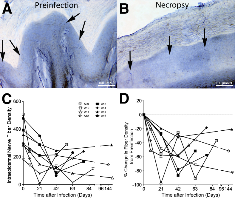
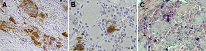
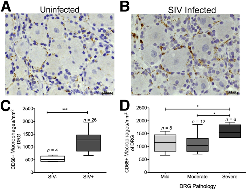
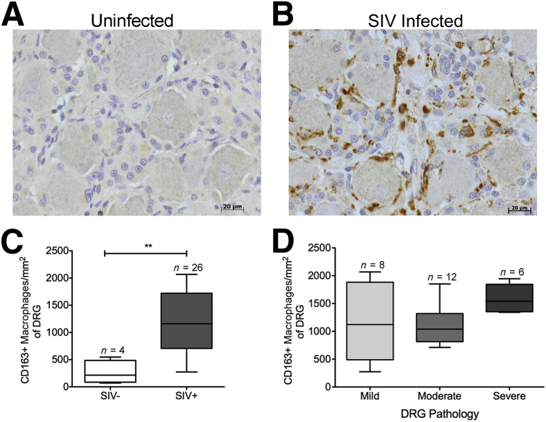
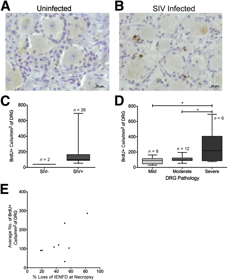
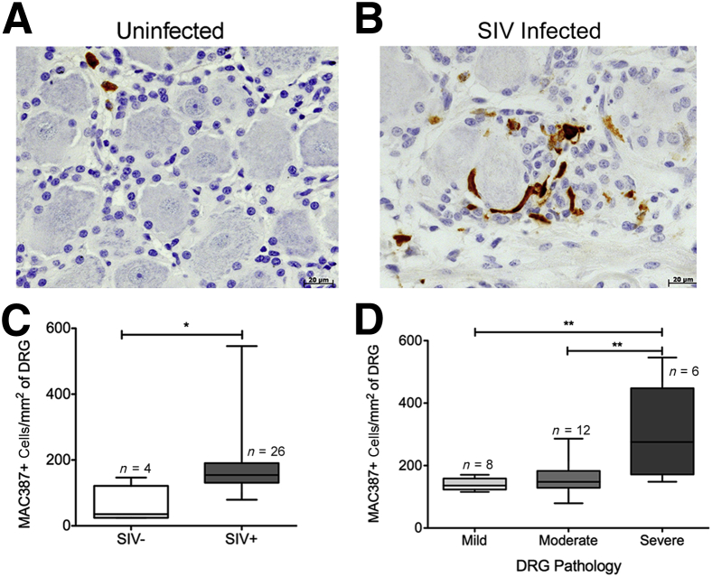
HIV-associated sensory neuropathy remains the most common neurological complication of HIV infection and is characterized by dorsal root ganglion (DRG) inflammation and intraepidermal nerve fiber density (IENFD) loss. Chronic peripheral immune cell activation and accumulation may cause damage to the DRG, but has not been fully investigated yet. By using an SIV-infected, CD8-lymphocyte-depleted rhesus macaque model, we defined immune cells surrounding DRG neurons and their role in DRG pathology, measured cell traffic from the bone marrow to the DRGs using 5-bromo-2-deoxyuridine (BrdU) pulse, and serially measured IENFD. We found an increase in CD68(+) and CD163(+) macrophages in DRGs of SIV-infected animals. MAC387(+) recently recruited monocytes/macrophages were increased, along with BrdU(+) cells, in the DRGs of SIV-infected macaques. We demonstrated that 78.1% of all BrdU(+) cells in DRGs were also MAC387(+). The number of BrdU(+) monocytes correlated with severe DRG histopathology, which included neuronophagia, neuronal loss, and Nageotte nodules. These data demonstrate that newly recruited MAC387(+)BrdU(+) macrophages may play a significant role in DRG pathogenesis. IENFD decreased early (day 21), consistent with the development of sensory neuropathy in SIV-infected macaques. Decreased IENFD was associated with elevated BrdU(+) cells in the DRG. These data suggest that increased recruitment of macrophages to DRG is associated with severe DRG histopathology and IENFD loss.


Figures

Figure 1
Figure 1
Intraepidermal nerve fiber density (IENFD) decreases after infection. A: Preinfection biopsy of animal A11 has several long contiguous IENFs terminating in the basement layer of the stratum corneum (arrows). B: Biopsy specimen of animal A11 taken at necropsy on AIDS has few segmented IENFs (arrows). C: IENFD was serially measured, and the IENFD per animal at each time point is graphed. D: The percentage change in IENFD at the individual times from preinfection was calculated using the animal's own preinfection value as a reference. Open symbols indicate cohort of SIV-infected animals (A09 to A12) in which biopsy specimens were taken at days 0, 8, 21, 42, and 63 after infection and necropsy; closed symbols, cohort of SIV-infected animals (A13 to A16) in which biopsy specimens were taken at days 0, 21, 42, and 63 after infection and necropsy.
Figure 2
Figure 2
Productive viral replication in the macrophage in the dorsal nerve root and dorsal root ganglion (DRG) of SIV-infected macaques. A: Dorsal nerve root of animal A11 with abundant SIVp28 (brown) immunoreactivity and multinucleated giant cells (MNGCs) in SIV-infected rhesus macaque. B: DRG of animal A06 with abundant SIVp28 (brown) immunoreactivity and a MNGC. C: DRG of animal A05 with in situ hybridization identifying SIV RNA+ cells (blue).
Figure 3
Figure 3
Elevated numbers of CD68+ macrophages are associated with SIV infection and severity of dorsal root ganglion (DRG) pathology. A: DRG of uninfected animal A01 with scant CD68 immunoreactivity (brown). B: DRG of animal A08, a SIV-infected rhesus macaque with marked increase in CD68 immunoreactivity (brown). C: The box plot shows the absolute number of CD68+ cells per mm2 in SIV and SIV+ DRGs. The absolute number of CD68+ cells per mm2 significantly increases in the SIV-infected DRGs. D: The box plot shows the absolute number of CD68+ cells per mm2 in mild, moderate, and severe DRGs. Elevated numbers of CD68+ macrophages are associated with severity of DRG pathology. Analysis of variance (P < 0.01) was performed, followed by post hoc t-tests. Data are given as means ± SEM (C and D). n = 4 (C, SIV DRGs); n = 26 (C, SIV+ DRGs); n = 8 (D, mild DRGs); n = 12 (D, moderate DRGs); n = 6 (D, severe DRGs). P < 0.05, ∗∗∗P < 0.001.
Figure 4
Figure 4
Elevated numbers of CD163+ macrophages are associated with SIV infection. A: Dorsal root ganglion (DRG) of uninfected animal A02 with scant CD163 immunoreactivity (brown). B: DRG of SIV-infected animal A05 with marked increase in CD163 immunoreactivity (brown). C: The box plot shows the absolute number of CD163+ cells per mm2 in SIV and SIV+ DRGs. The absolute number of CD163+ cells per mm2 significantly increases in the SIV-infected group. D: The box plot shows the absolute number of CD163+ cells per mm2 in mild, moderate, and severe DRGs. Elevated numbers of CD163+ macrophages were not associated with severity of DRG pathology. Data are given as means ± SEM (C and D). n = 4 (C, SIV DRGs); n = 26 (C, SIV+ DRGs); n = 8 (D, mild DRGs); n = 12 (D, moderate DRGs); n = 6 (D, severe DRGs). ∗∗P < 0.01.
Figure 5
Figure 5
Cell traffic from the bone marrow to the dorsal root ganglion (DRG) measured by increased 5-bromo-2-deoxyuridine (BrdU+) cells with SIV infection. Animals were serially injected with BrdU to label recently divided monocytes in the bone marrow and then traffic to DRG. A: DRG of uninfected animal A02 with scant BrdU immunoreactivity (brown). B: DRG of SIV-infected animal A06 with marked increase in BrdU immunoreactivity (brown). C: The box plot shows the absolute number of BrdU+ cells per mm2 in SIV and SIV+ DRGs. The absolute number of BrdU+ cells per mm2 of DRG tissue was calculated. D: The box plot shows the absolute number of BrdU+ cells per mm2 in mild, moderate, and severe DRGs. Higher numbers of BrdU+ cells correlate with the severity of DRG pathology. Analysis of variance (P < 0.01) was performed, followed by post hoc t-tests. E: The average number of BrdU+ cells in the DRGs per animal was calculated. Increased numbers of BrdU+ cells in the DRG correlate with percentage loss of intraepidermal nerve fiber density (IENFD) at necropsy. Spearman correlation was used. Data are given as means ± SEM (C and D). n = 4 (C, SIV DRGs); n = 26 (C, SIV+ DRGs); n = 8 (D, mild DRGs); n = 12 (D, moderate DRGs); n = 6 (D, severe DRGs). P < 0.05 (r = 0.064).
Figure 6
Figure 6
Elevated numbers of MAC387+ macrophages are associated with SIV infection and severity of dorsal root ganglion (DRG) pathology. A: DRG of uninfected animal A01 with scant MAC387 immunoreactivity (brown). B: DRG of SIV-infected animal A05 with marked increase in MAC387 immunoreactivity (brown). C: The box plot shows the absolute number of MAC387+ cells per mm2 in SIV and SIV+ DRGs. The absolute number of MAC387+ cells per mm2 significantly increases in the SIV-infected group. D: The box plot shows the absolute number of MAC387+ cells per mm2 in mild, moderate, and severe DRGs. Elevated number of MAC387+ macrophages is associated with severity of DRG pathology. Analysis of variance (P < 0.01) was performed, followed by post hoc t-tests. Data are given as means ± SEM (C and D). n = 4 (C, SIV DRGs); n = 26 (C, SIV+ DRGs); n = 8 (D, mild DRGs); n = 12 (D, moderate DRGs); n = 6 (D, severe DRGs). P < 0.05, ∗∗P < 0.01.

References

    1. Ellis R.J., Rosario D., Clifford D.B., McArthur J.C., Simpson D., Alexander T., Gelman B.B., Vaida F., Collier A., Marra C.M., Ances B., Atkinson J.H., Dworkin R.H., Morgello S., Grant I. Continued high prevalence and adverse clinical impact of human immunodeficiency virus-associated sensory neuropathy in the era of combination antiretroviral therapy: the CHARTER Study. Arch Neurol. 2010;67:552–558. - PMC - PubMed
    1. Nicholas P.K., Mauceri L., Slate Ciampa A., Corless I.B., Raymond N., Barry D.J., Viamonte Ros A. Distal sensory polyneuropathy in the context of HIV/AIDS. J Assoc Nurses AIDS Care. 2007;18:32–40. - PubMed
    1. Verma S., Estanislao L., Simpson D. HIV-associated neuropathic pain: epidemiology, pathophysiology and management. CNS Drugs. 2005;19:325–334. - PubMed
    1. Ellis R.J., Marquie-Beck J., Delaney P., Alexander T., Clifford D.B., McArthur J.C., Simpson D.M., Ake C., Collier A.C., Gelman B.B., McCutchan J.A., Morgello S., Grant I. Human immunodeficiency virus protease inhibitors and risk for peripheral neuropathy. Ann Neurol. 2008;64:566–572. - PMC - PubMed
    1. Pardo C.A., McArthur J.C., Griffin J.W. HIV neuropathy: insights in the pathology of HIV peripheral nerve disease. J Peripher Nerv Syst. 2001;6:21–27. - PubMed

Publication types

MeSH terms