

Elevated intraocular pressure induces Rho GTPase mediated contractile signaling in the trabecular meshwork
- PMID: 25956210
- PMCID: PMC4466129
- DOI: 10.1016/j.exer.2015.05.001
Elevated intraocular pressure induces Rho GTPase mediated contractile signaling in the trabecular meshwork
Abstract
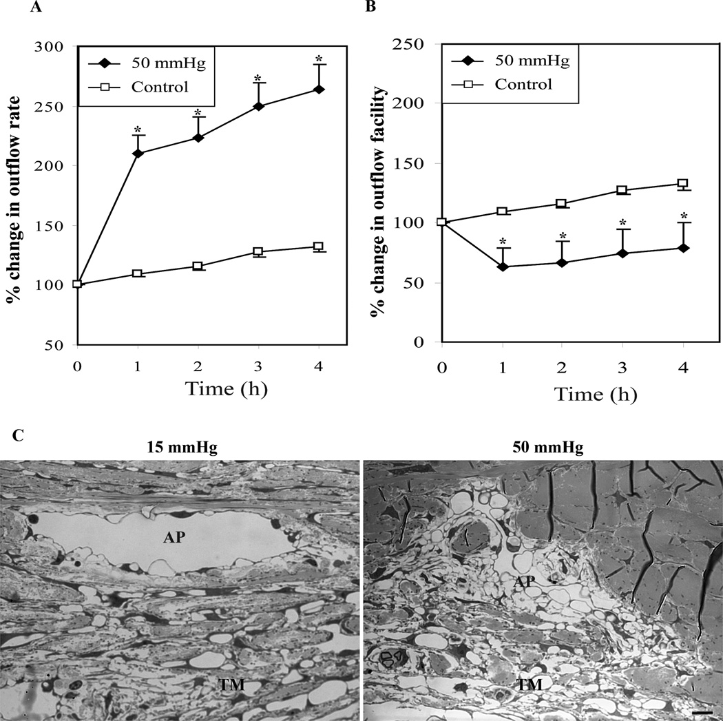
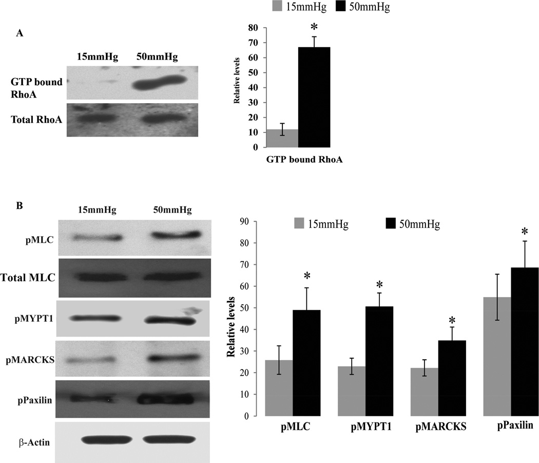
Rho GTPase regulated contractile signaling in the trabecular meshwork (TM) has been shown to modulate aqueous humor (AH) outflow and intraocular pressure (IOP). To explore whether elevated IOP, a major risk factor for primary open angle glaucoma (POAG) influences Rho GTPase signaling in the TM, we recorded AH outflow in enucleated contralateral porcine eyes perfused for 4-5 h at either 15 mm or 50 mm Hg pressure. After perfusion, TM tissue extracted from perfused eyes was evaluated for the activation status of Rho GTPase, myosin light chain (MLC), myosin phosphatase target substrate 1 (MYPT1), myristoylated alanine-rich C-kinase substrate (MARCKS) and paxillin. Eyes perfused at 50 mm Hg exhibited a significant decrease in AH outflow facility compared with those perfused at 15 mm Hg. Additionally, TM tissue from eyes perfused at 50 mm Hg revealed significantly increased levels of activated RhoA and phosphorylated MLC, MYPT1, MARCKS and paxillin compared to TM tissue derived from eyes perfused at 15 mm Hg. Taken together, these observations indicate that elevated IOP-induced activation of Rho GTPase-dependent contractile signaling in the TM is associated with increased resistance to AH outflow through the trabecular pathway, and demonstrate the sensitivity of Rho GTPase signaling to mechanical force in the AH outflow pathway.
Keywords: Intraocular pressure; Mechanotransduction; Outflow resistance; Rho GTPase; Trabecular meshwork.
Copyright © 2015 Elsevier Ltd. All rights reserved.
Conflict of interest statement
Figures
References
-
- Acott TS, Kelley MJ, Keller KE, Vranka JA, Abu-Hassan DW, Li X, Aga M, Bradley JM. Intraocular pressure homeostasis: maintaining balance in a high-pressure environment. Journal of ocular pharmacology and therapeutics : the official journal of the Association for Ocular Pharmacology and Therapeutics. 2014;30:94–101. - PMC - PubMed
-
- Aderem A. The MARCKS family of protein kinase-C substrates. Biochemical Society transactions. 1995;23:587–591. - PubMed
-
- Battista SA, Lu Z, Hofmann S, Freddo T, Overby DR, Gong H. Reduction of the available area for aqueous humor outflow and increase in meshwork herniations into collector channels following acute IOP elevation in bovine eyes. Investigative ophthalmology & visual science. 2008;49:5346–5352. - PMC - PubMed
-
- Becker B, Constant MA. The facility of aqueous outflow; a comparison of tonography and perfusion measurements in vivo and in vitro. AMA Arch Ophthalmol. 1956;55:305–312. - PubMed
Publication types
MeSH terms
Substances
Grants and funding
LinkOut - more resources
Full Text Sources
Other Literature Sources
Miscellaneous
