Review
. 2015 Sep;17(5):1041-54.
doi: 10.1208/s12248-015-9780-2. Epub 2015 May 9.

A Review of Clinical Translation of Inorganic Nanoparticles

Affiliations
Review

A Review of Clinical Translation of Inorganic Nanoparticles

Aaron C Anselmo et al. AAPS J. 2015 Sep.

Abstract

Inorganic nanoparticles are widely used for therapeutic and diagnostic purposes as they offer unique features as compared with their organic and polymeric counterparts. As such, inorganic nanoparticles represent an exciting opportunity to develop drug delivery and imaging systems that are poised to tackle unique challenges which are currently unaddressed in clinical settings. Despite these clear advantages, very few inorganic nanoparticle systems have entered the clinic. Here, we review the current clinical landscape of inorganic nanoparticle systems and their opportunities and challenges, with particular emphasis on gold-, iron-oxide- and silica-based nanoparticle systems. Key examples of inorganic nanoparticles that are currently being investigated in the clinic (e.g., trials which are recruiting or currently active but not completed) are highlighted, along with the preclinical work that these examples have leveraged to transition from the lab to the clinic.


Figures
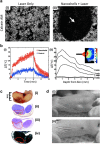
Fig. 1
Fig. 1
Preclinical performance of AuroLase® AuNPs currently undergoing clinical trials. a Membrane-viability-stained (calcein AM) breast cancer cells following laser exposure i without nanoshells and ii with nanoshells. The arrow in (ii) represents the area of dead cells that were exposed to both nanoshells and the laser. b Temperature change following NIR-irradiation of nanoshells in vivo: i as a function of time (nanoshells group: red circles, control group with no nanoshells: blue circles) and ii as a function of tissue depth (inset, heat map). c Antitumor effect of NIR-stimulated nanoshells treatment: i gross tumor images showing hemorrhaging, ii nanoshell localization (red outline) following silver staining, iii tissue damage as shown by hematoxylin and eosin staining, and iv magnetic resonance temperature imaging suggesting areas of irreversible thermal damage. d Prostate tumor regressions following a single treatment of an intravenous: i saline injection with laser treatment or ii nanoshell injection with laser treatment. ac Adapted from (29), copyright (2003) National Academy of Sciences, U.S.A. d Adapted from (31), copyright (2008), with permission from Elsevier
Fig. 2
Fig. 2
Preclinical and clinical performance of Sebashell AuNPs currently undergoing clinical trials. a NIR light absorption of Sebashells produces heat. b Schematic illustrating the topical application of Sebashells, ultrasound-mediated penetration of Sebashells, and NIR-laser-induced ablation of sebaceous glands using Sebashells. c Two-photon-induced photoluminescence imaging shows the localization of Sebashells in a sebaceous gland in porcine skin in vitro. d Deep glandular thermolysis following NIR-induced ablation of sebaceous gland localized Sebashells. e Clinical data in humans showing thermolysis as a function of ultrasound exposure. ae Adapted from (39), copyright (2015), with permission from Elsevier
Fig. 3
Fig. 3
Clinical pathological imaging using ferumoxytol nanoparticles. MRI of patient with a known glioblastoma multiforme. T1-weighted MRI images: a precontrast, b gadoteridol enhanced, and c 24-h post-ferumoxytol enhanced. T2-weighted MRI images: d precontrast, e gadoteridol enhanced, and f 24-h post-ferumoxytol enhanced. White arrows indicate enhancement regions present in ferumoxytol groups but not in gadoteridol groups. g Ferumoxytol MRI in a representative patient with type-1 diabetes (left) and a non-diabetic control patient (right). Single slices (top) and 3D volumes (bottom) show increased ferumoxytol accumulation in a diabetic patient as compared with control. af Reprinted from (69), by permission of Oxford University Press. g Adapted from (71)
Fig. 4
Fig. 4
Preclinical and clinical performance of C-Dots. a In vivo fluorescent imaging in nude mice, 45 min after injection, with: i bare C-Dots, which show accumulation in liver and bladder, and ii PEG-coated C-Dots, which show accumulation only in bladder. PEG coating effectively allows C-Dots to avoid reticuloendothelial clearance organs, such as the liver, and eventually accumulate in the bladder prior to renal clearance. b C-Dot schematic showing: integrin-targeting cRGDY functionality, 124I-labeling to facilitate PET imaging, Cy5-loaded core to facilitate fluorescent imaging and PEG coating to allow immune system avoidance. c i Coronal CT showing left hepatic lobe metastasis (yellow arrowhead), ii coronal PET image, 4-h post-C-Dot injection, showing particle accumulation around tumor periphery (yellow arrowhead), and iii co-registered PET-CT, 4-h post-C-Dot injection, highlighted C-Dot accumulation in tumor periphery in a patient with anorectal mucosa melanoma, with liver metastasis. d i MRI, 72-h post-C-Dot injection, showing a cystic focus in the anterior pituitary gland, ii PET-CT imaging showing C-Dot accumulation in pituitary lesion, and iii MRI-PET showing overlap of C-Dots from PET with pituitary lesion from MRI in a patient with a pituitary microadenoma. iv Time-lapse PET imaging highlighting the progressive accumulation of C-Dot activity up to 72-h post-injection. e In a patient with impaired renal function and chemotherapy related nephrotoxicity, PET imaging showed C-Dot activity in the cardiac blood pool (yellow arrow), small bowel (black arrowhead), renal cortices (red arrows), and bladder (asterisk); 72 h later, C-Dot activity remains in the renal cortices. a Adapted with permission from (93). Copyright (2009) American Chemical Society. be From (89). Adapted with permission from AAAS

References

    1. Huang H-C, Barua S, Sharma G, Dey SK, Rege K. Inorganic nanoparticles for cancer imaging and therapy. J Control Release. 2011;155(3):344–57. doi: 10.1016/j.jconrel.2011.06.004. - DOI - PubMed
    1. Na HB, Song IC, Hyeon T. Inorganic nanoparticles for MRI contrast agents. Adv Mater. 2009;21(21):2133–48. doi: 10.1002/adma.200802366. - DOI
    1. Li Z, Barnes JC, Bosoy A, Stoddart JF, Zink JI. Mesoporous silica nanoparticles in biomedical applications. Chem Soc Rev. 2012;41(7):2590–605. doi: 10.1039/c1cs15246g. - DOI - PubMed
    1. Giljohann DA, Seferos DS, Daniel WL, Massich MD, Patel PC, Mirkin CA. Gold nanoparticles for biology and medicine. Angew Chem Int Ed. 2010;49(19):3280–94. doi: 10.1002/anie.200904359. - DOI - PMC - PubMed
    1. McCarthy JR, Weissleder R. Multifunctional magnetic nanoparticles for targeted imaging and therapy. Adv Drug Deliv Rev. 2008;60(11):1241–51. doi: 10.1016/j.addr.2008.03.014. - DOI - PMC - PubMed

Publication types