

Geometric Calibration and Image Reconstruction for a Segmented Slant-Hole Stationary Cardiac SPECT System
- PMID: 25956691
- PMCID: PMC5297456
- DOI: 10.2967/jnmt.114.153668
Geometric Calibration and Image Reconstruction for a Segmented Slant-Hole Stationary Cardiac SPECT System
Abstract
A dedicated stationary cardiac single-photon emission computed tomography (SPECT) system with a novel segmented slant-hole collimator has been developed. The goal of this paper is to calibrate this new imaging geometry with a point source.
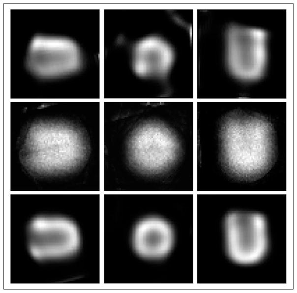
Methods: Unlike the commercially available dedicated cardiac SPECT systems, which are specialized and can be used only to image the heart, our proposed cardiac system is based on a conventional SPECT system but with a segmented slant-hole collimator replacing the collimator. For a dual-head SPECT system, 2 segmented collimators, each with 7 sections, are arranged in an L-shaped configuration such that they can produce a complete cardiac SPECT image with only one gantry position. A calibration method was developed to estimate the geometric parameters of each collimator section as well as the detector rotation radius, under the assumption that the point source location is calculated using the central-section data. With a point source located off the rotation axis, geometric parameters for each collimator section can be estimated independently. The parameters estimated individually are further improved by a joint objective function that uses all collimator sections simultaneously and incorporates the collimator symmetry information.
Results: Estimation results and images reconstructed from estimated parameters are presented for both simulated and real data acquired from a prototype collimator. The calibration accuracy was validated by computer simulations with an error of about 0.1° for the slant angles and about 1 mm for the rotation radius. Reconstructions of a heart-insert phantom did not show any image artifacts of inaccurate geometric parameters.
Conclusion: Compared with the detector's intrinsic resolution, the estimation error is small and can be ignored. Therefore, the accuracy of the calibration is sufficient for cardiac SPECT imaging.
Keywords: cardiac single photon emission computed tomography (SPECT); collimator; geometric calibration; slant-hole.
© 2015 by the Society of Nuclear Medicine and Molecular Imaging, Inc.
Figures


References
-
- Babla H, Bai C, Conwell R. A triple-head solid state camera for cardiac single photon emission tomography (SPECT) Proc SPIE. 2006;6319:63190M.1–63190M.5.
-
- Funk T, Kirch DL, Koss JE, Botvinick E, Hasegawa BH. A novel approach to multipinhole SPECT for myocardial perfusion imaging. J Nucl Med. 2006;47:595–602. - PubMed
-
- Steele PP, Kirch DL, Koss JE. Comparison of simultaneous dual-isotope multi-pinhole SPECT with rotational SPECT in a group of patients with coronary artery disease. J Nucl Med. 2008;49:1080–1089. - PubMed
-
- Erlandsson K, Kacperski K, Van Gramberg D, Hutton BF. Performance evaluation of D-SPECT: a novel SPECT system for nuclear cardiology. Phys Med Biol. 2009;54:2635–2649. - PubMed
Publication types
MeSH terms
Grants and funding
LinkOut - more resources
Full Text Sources
Other Literature Sources