. 2015 Jul;81(14):4756-66.
doi: 10.1128/AEM.00772-15. Epub 2015 May 8.

Stoichiometric Assembly of the Cellulosome Generates Maximum Synergy for the Degradation of Crystalline Cellulose, as Revealed by In Vitro Reconstitution of the Clostridium thermocellum Cellulosome

Affiliations

Stoichiometric Assembly of the Cellulosome Generates Maximum Synergy for the Degradation of Crystalline Cellulose, as Revealed by In Vitro Reconstitution of the Clostridium thermocellum Cellulosome

Katsuaki Hirano et al. Appl Environ Microbiol. 2015 Jul.

Abstract

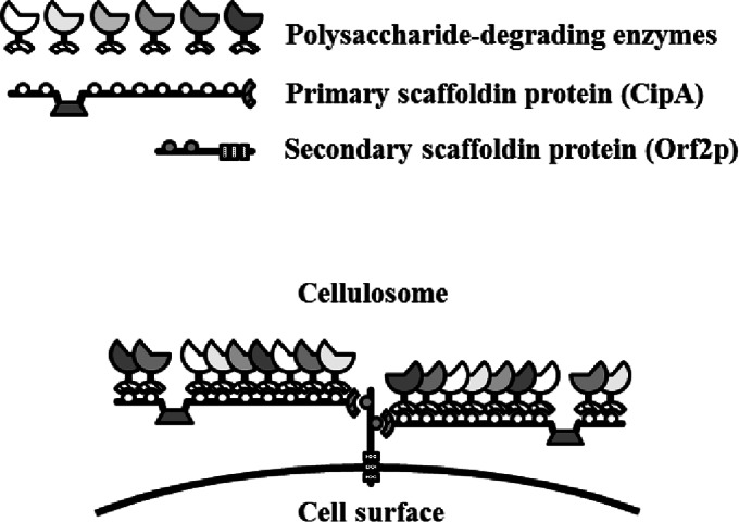
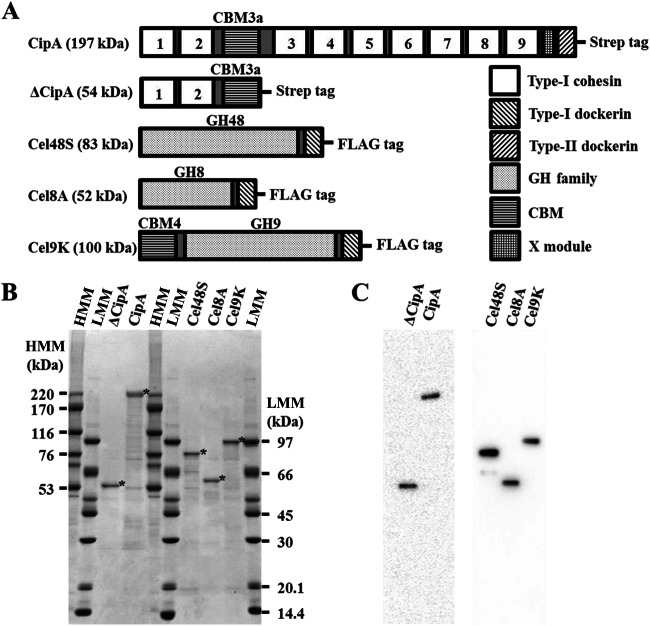
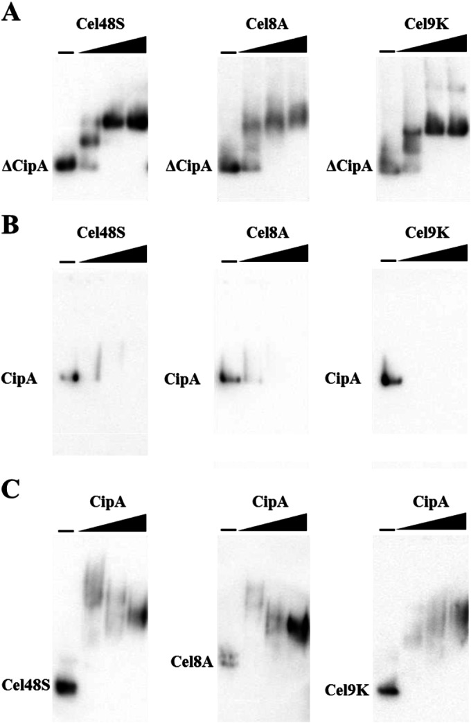
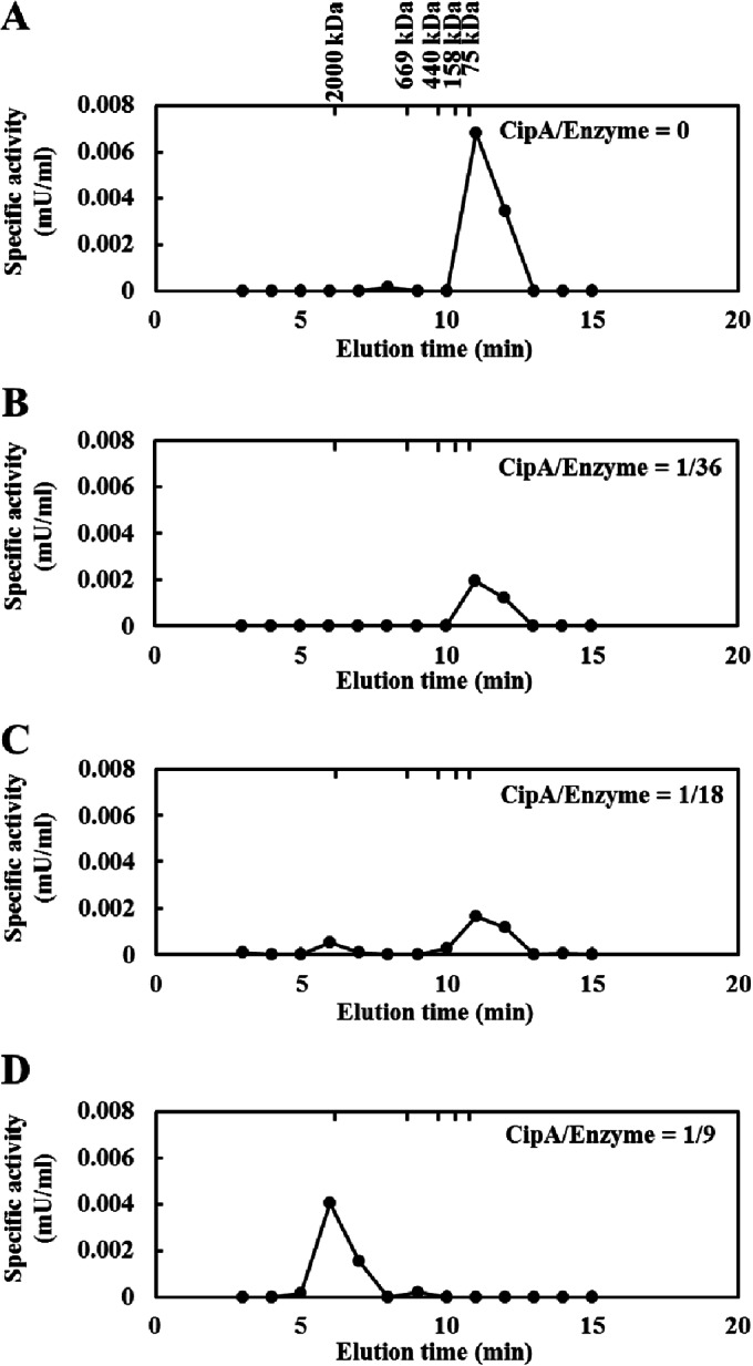
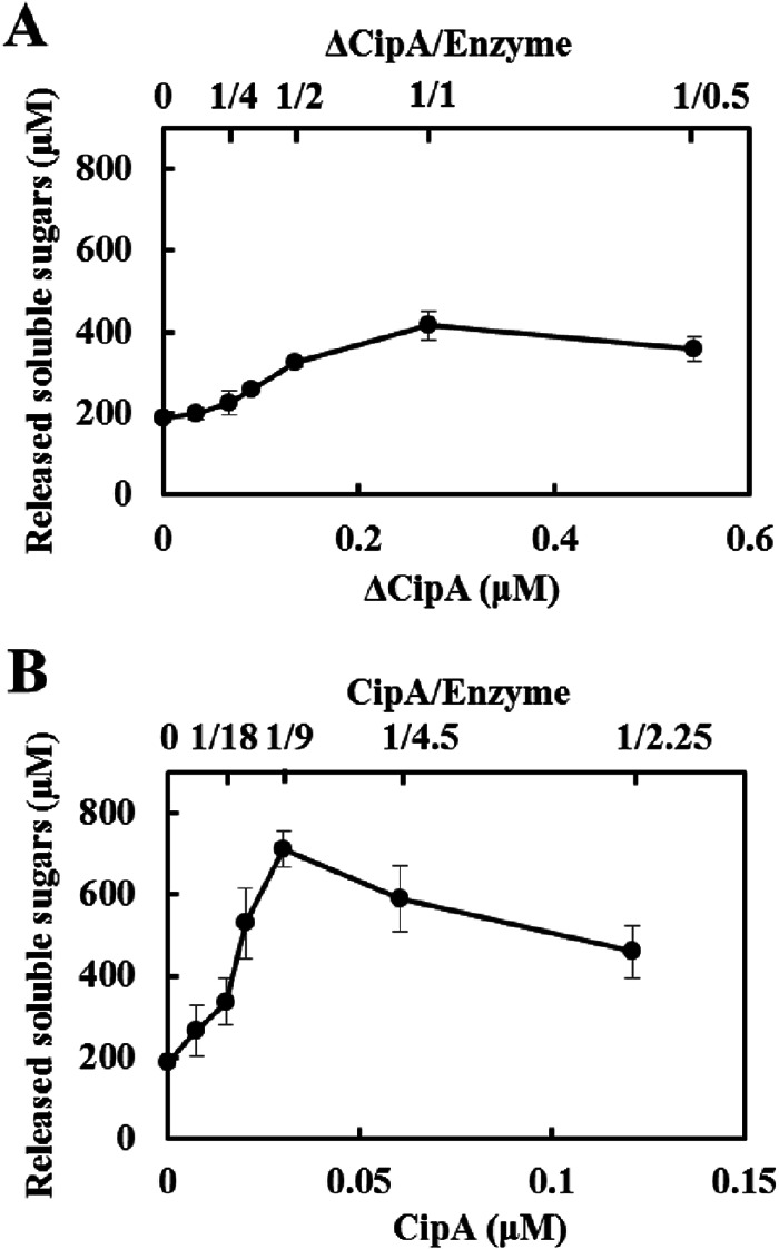
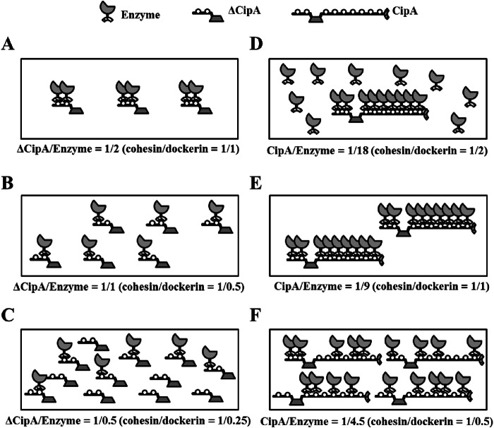
The cellulosome is a supramolecular multienzyme complex formed by species-specific interactions between the cohesin modules of scaffoldin proteins and the dockerin modules of a wide variety of polysaccharide-degrading enzymes. Cellulosomal enzymes bound to the scaffoldin protein act synergistically to degrade crystalline cellulose. However, there have been few attempts to reconstitute intact cellulosomes due to the difficulty of heterologously expressing full-length scaffoldin proteins. We describe the synthesis of a full-length scaffoldin protein containing nine cohesin modules, CipA; its deletion derivative containing two cohesin modules, ΔCipA; and three major cellulosomal cellulases, Cel48S, Cel8A, and Cel9K, of the Clostridium thermocellum cellulosome. The proteins were synthesized using a wheat germ cell-free protein synthesis system, and the purified proteins were used to reconstitute cellulosomes. Analysis of the cellulosome assembly using size exclusion chromatography suggested that the dockerin module of the enzymes stoichiometrically bound to the cohesin modules of the scaffoldin protein. The activity profile of the reconstituted cellulosomes indicated that cellulosomes assembled at a CipA/enzyme molar ratio of 1/9 (cohesin/dockerin = 1/1) and showed maximum synergy (4-fold synergy) for the degradation of crystalline substrate and ∼2.4-fold-higher synergy for its degradation than minicellulosomes assembled at a ΔCipA/enzyme molar ratio of 1/2 (cohesin/dockerin = 1/1). These results suggest that the binding of more enzyme molecules on a single scaffoldin protein results in higher synergy for the degradation of crystalline cellulose and that the stoichiometric assembly of the cellulosome, without excess or insufficient enzyme, is crucial for generating maximum synergy for the degradation of crystalline cellulose.


Figures

FIG 1
FIG 1
Schematic representation of the C. thermocellum cellulosome. The cellulosome is a cell surface-displayed supramolecular multienzyme complex containing a wide variety of polysaccharide-degrading enzymes. The formation of the C. thermocellum cellulosome is mediated by two specific interactions. One interaction is between the type I dockerin module at the C termini of polysaccharide-degrading enzymes and the internal nine cohesin modules of the primary scaffoldin protein, and the other interaction is mediated between the type II dockerin module at the C terminus of the primary scaffoldin protein and the internal cohesin modules of the cell surface-displayed secondary scaffoldin protein. The scaffold of the cellulosome complex assembles through the interaction of one primary scaffoldin protein (CipA, containing nine type I cohesin modules) and four secondary scaffoldin proteins (SdbA, which contains one type II cohesin module; Orf2p, which contains two type II cohesin modules; OlpB, which contains seven type II cohesin modules; and Cthe0736, which contains seven type II cohesin modules). In the diagram, Orf2p is used as a representative secondary scaffoldin protein.
FIG 2
FIG 2
Domain organization and SDS-PAGE analysis of the cellulosomal components. (A) The C. thermocellum cellulosomal scaffoldin protein, CipA, contains nine type I cohesin modules (cohesins 1 to 9), a CBM3a, an X module, and a type II dockerin module. The N-terminal segment of CipA, designated ΔCipA, contains two type I cohesin modules (cohesins 1 and 2) and a CBM3a. The model complex employed here contained three enzymatic components: Cel48S, a GH48 enzyme; Cel8A, a GH8 enzyme; and Cel9K, a GH9 enzyme that also contains a CBM4 module at its N terminus. All three enzymes contain a type I dockerin module at their C termini. The dockerin module specifically associates with the type I cohesin modules of the scaffoldin protein. Mature scaffoldin and enzymatic components were synthesized in a cell-free system as GST fusion proteins containing a Strep and a FLAG tag, respectively, at the C terminus and then purified by cleavage with PreScission protease in a column. (B) Purified samples were subjected to SDS-PAGE on a 4 to 20% gel and stained with Coomassie brilliant blue. The bands corresponding to the purified proteins are indicated by asterisks. HMM, high molecular mass; LMM, low molecular mass. (C) Purified proteins were detected by Western blotting with anti-Strep or anti-FLAG M2 monoclonal antibody that targeted the C-terminal Strep or FLAG tag of the scaffoldin or enzymatic component, respectively.
FIG 3
FIG 3
Electrophoretic mobility shift assay of the minicellulosome and cellulosome assembly. (A) Minicellulosome complexes assembled by mixing ΔCipA at a fixed concentration (0.050 μM) and each enzyme at various concentrations. The molar ratio of ΔCipA containing two cohesin modules to the enzyme containing one dockerin module is designated “ΔCipA/enzyme,” and the molar ratio of the cohesin module to the dockerin module is designated “cohesin/dockerin.” The complexes were assembled at ΔCipA/enzyme molar ratios of 1/0, 1/1, 1/2, and 1/4 (cohesin/dockerin = 1/0, 1/0.5, 1/1, and 1/2) and analyzed by native PAGE on 4 to 20% gradient gels with Western blot analysis using an anti-Strep tag monoclonal antibody. The bands corresponding to ΔCipA are indicated. (B) Cellulosome complexes assembled by mixing CipA at a fixed concentration (0.025 μM) and each enzyme at various concentrations. The molar ratio of CipA containing nine cohesin modules to the enzyme containing one dockerin module is designated “CipA/enzyme.” The complexes were assembled at CipA/enzyme molar ratios of 1/0, 1/4.5, 1/9, and 1/18 (cohesin/dockerin = 1/0, 1/0.5, 1/1, and 1/2) and detected by Western blotting with an anti-Strep tag monoclonal antibody. The bands corresponding to CipA are indicated. (C) Cellulosome complexes assembled by mixing Cel48S, Cel8A, or Cel9K at a fixed concentration (0.050 μM) and CipA at various concentrations. The complexes were assembled at CipA/enzyme molar ratios of 0, 1/36, 1/9, and 1/2.25 (cohesin/dockerin = 0, 1/4, 1/1, and 1/0.25) and detected by Western blotting with anti-FLAG M2 tag monoclonal antibody. The bands corresponding to Cel48S, Cel8A, and Cel9K are indicated.
FIG 4
FIG 4
Size exclusion chromatography of reconstituted cellulosome complexes. Stoichiometric assembly of the reconstituted cellulosome complex was analyzed by size exclusion chromatography as described in Materials and Methods. The cellulosome complexes were assembled by mixing CipA at various concentrations with the enzyme mixture at a fixed concentration (0.41 μM). The enzyme mixture comprised a Cel48S/Cel8A/Cel9K molar ratio of 4.06:1.82:0.72. The CipA/enzyme values indicate the molar ratio of CipA to the enzyme mixture; for example, a cellulosome complex assembled with 0.045 μM CipA and 0.41 μM enzyme mixture is shown as “CipA/enzyme = 1/9.” The complexes were assembled at CipA/enzyme molar ratios of 0 (cohesin/dockerin = 0) (A), 1/36 (cohesin/dockerin = 1/4) (B), 1/18 (cohesion/dockerin = 1/2) (C), and 1/9 (cohesin/dockerin = 1/1) (D). The enzymatic activity was determined by measuring the amount of reducing sugars released from 0.5% PASC. The molecular mass markers (2,000-kDa Blue Dextran, 669-kDa thyroglobulin, 440-kDa ferritin, 158-kDa aldolase, and 75-kDa conalbumin) are indicated.
FIG 5
FIG 5
Enzymatic activities of reconstituted minicellulosome and cellulosome complexes for crystalline cellulose. The enzymatic activities of minicellulosome (A) and cellulosome (B) complexes were determined by measuring the amount of reducing sugars released from 0.5% Avicel at 55°C in 24 h. Minicellulosome or cellulosome complexes were assembled by mixing ΔCipA or CipA at various concentrations with the enzyme mixture at a fixed concentration (0.27 μM). The enzyme mixture comprised a Cel48S/Cel8A/Cel9K molar ratio of 4.06:1.82:0.72. The scaffold/enzyme values indicate the molar ratio of scaffoldin protein to the enzyme mixture; for example, a cellulosome complex assembled with 0.030 μM CipA and 0.27 μM enzyme mixture is indicated as 1/9. The data are presented as the means from three independent experiments ± standard errors (SE).
FIG 6
FIG 6
Schematic models of minicellulosome and cellulosome complexes. The minicellulosome or cellulosome complexes were assembled by mixing ΔCipA or CipA at various concentrations with an enzyme mixture at a fixed concentration. (A) The minicellulosome complex assembled at a ΔCipA/enzyme ratio of 1/2 (cohesin/dockerin = 1/1) is predicted to comprise an enzyme-saturated complex displaying two enzyme molecules. (B and C) The complexes assembled at ΔCipA/enzyme ratios of 1/1 (cohesin/dockerin = 1/0.5) (B) and 1/0.5 (cohesin/dockerin = 1/0.25) (C) are predicted to display only one enzyme molecule. (D) In contrast, the cellulosome complex assembled at a CipA/enzyme ratio of 1/18 (cohesin/dockerin = 1/2) is predicted to comprise an enzyme-saturated complex displaying nine enzyme molecules and the free enzymes. (E) The complex assembled at a CipA/enzyme ratio of 1/9 (cohesin/dockerin = 1/1) is predicted to comprise the enzyme-saturated complex. (F) The complex assembled at a CipA/enzyme ratio of 1/4.5 (cohesin/dockerin = 1/0.5) is predicted to display fewer than nine enzyme molecules.

Similar articles

Cited by

References

    1. Bayer EA, Belaich JP, Shoham Y, Lamed R. 2004. The cellulosomes: multienzyme machines for degradation of plant cell wall polysaccharides. Annu Rev Microbiol 58:521–554. doi:10.1146/annurev.micro.57.030502.091022. - DOI - PubMed
    1. Demain AL, Newcomb M, Wu JH. 2005. Cellulase, clostridia, and ethanol. Microbiol Mol Biol Rev 69:124–154. doi:10.1128/MMBR.69.1.124-154.2005. - DOI - PMC - PubMed
    1. Gold ND, Martin VJ. 2007. Global view of the Clostridium thermocellum cellulosome revealed by quantitative proteomic analysis. J Bacteriol 189:6787–6795. doi:10.1128/JB.00882-07. - DOI - PMC - PubMed
    1. Raman B, Chongle P, Hurst GB, Rodriguez M, McKeown CM, Lankford PK, Samatova NF, Mielenz JR. 2009. Impact of pretreated switchgrass and biomass carbohydrates on Clostridium thermocellum ATCC 27405 cellulosome composition: a quantitative proteomic analysis. PLoS One 4:e5271. doi:10.1371/journal.pone.0005271. - DOI - PMC - PubMed
    1. Kruus K, Wang WK, Ching J, Wu JH. 1995. Exoglucanase activities of the recombinant Clostridium thermocellum CelS, a major cellulosome component. J Bacteriol 177:1641–1644. - PMC - PubMed

Publication types

MeSH terms

LinkOut - more resources