Review
. 2016 Jan;275 Pt 2(0 2):245-52.
doi: 10.1016/j.expneurol.2015.04.022. Epub 2015 May 6.

Neonatal sensory nerve injury-induced synaptic plasticity in the trigeminal principal sensory nucleus

Affiliations
Review

Neonatal sensory nerve injury-induced synaptic plasticity in the trigeminal principal sensory nucleus

Fu-Sun Lo et al. Exp Neurol. 2016 Jan.

Abstract

Sensory deprivation studies in neonatal mammals, such as monocular eye closure, whisker trimming, and chemical blockade of the olfactory epithelium have revealed the importance of sensory inputs in brain wiring during distinct critical periods. But very few studies have paid attention to the effects of neonatal peripheral sensory nerve damage on synaptic wiring of the central nervous system (CNS) circuits. Peripheral somatosensory nerves differ from other special sensory afferents in that they are more prone to crush or severance because of their locations in the body. Unlike the visual and auditory afferents, these nerves show regenerative capabilities after damage. Uniquely, damage to a somatosensory peripheral nerve does not only block activity incoming from the sensory receptors but also mediates injury-induced neuro- and glial chemical signals to the brain through the uninjured central axons of the primary sensory neurons. These chemical signals can have both far more and longer lasting effects than sensory blockade alone. Here we review studies which focus on the consequences of neonatal peripheral sensory nerve damage in the principal sensory nucleus of the brainstem trigeminal complex.

Keywords: Astrocytes; Infraorbital nerve; Reactive synaptogenesis; Silent synapses; Whisker–barrel system.


Conflict of interest statement

The authors declare no conflict of interest.

Figures

Figure 1
Figure 1. Barrelettes and the physiological properties of PrV neurons
A. PrV appears as a peanut-shaped structure in coronal sections through the brainstem. The five rows of whiskers on the face are represented in an inverted fashion in the PrV with the dorsal most whiskers represented ventrally and the tip of the nose medially. The nucleus is bordered dorsomedially by the motor trigeminal nucleus and laterally by the central trigeminal tract (TrV); d: dorsal, l; lateral. Cytochrome oxidase histochemistry reveals the barrelettes patterns in the ventral half of the nucleus. B. Schematic representation of the barrelettes as cellular modules (top) and cytochrome oxidase dense patches (bottom). C. Three types of PrV cells in the barrelettes region. The drawings illustrate barrelettes (dark blue) interbarrelette (light blue) and GABAergic interneurons (red). Whole-cell recording in barrelette neuron shows A-type potassium conductance upon membrane depolarization (dark blue record). The same membrane depolarization induces a low threshold spike mediated by T-type calcium channels (light blue record) in interbarrelette neuron. GABAergic neuron gives fast spiking firing to the membrane depolarization (red record).
Figure 2
Figure 2. Neuronal circuitry in the PrV
Activation of trigeminal ION central axon S1 induces a monosynaptic EPSP and a disynaptic feed-forward IPSP in the recorded barrelette neuron (dark blue) via GABAergic interneuron (red). Activation of S2 gives rise to a disynaptic lateral IPSP alone in the same barrelette neuron. Stimulation of both S1 and S2 with increasing intensity results in stepwise amplitude increase in both EPSP and IPSP. However, the step number of IPSC is larger than that of EPSP, suggesting the GABergic interneuron receives trigeminal inputs from more fibers than the recorded barrelette neuron, i.e., both feed-forward and lateral inhibition are mediated by the same group GABAergic interneurons. Similarly, the step number of EPSP in interbarrelette neuron (light blue) is larger than that of barrelette neuron, suggesting that interbarrelette neuron receives more trigeminal inputs than barrelette neuron.
Figure 3
Figure 3. Effects of neonatal ION damage in the PrV
A. Illustration of the organization of whiskers and corresponding barrelettes in the PrV. B. A similar illustration depicting the effects of IO nerve cut or nerve crush on barrelettes, there are no longer whisker-related patterns. C, D, Cyrochrome oxidase histochemistry reveals barrelettes in postnatal PrV (C) and their absence following ION damage (D). Scale bar = 300 um. E, F, similar micrographs showing vesicular glutamate 1 (VGlut1) immunolabeling showing how trigeminal afferents in the ION projection zone of the PrV no longer show immunoreactivity after neonatal nerve damage. VGlut1 is used as a marker for trigeminal afferent axons and their terminals. The immunoreactive patches seen at the very ventral part of the PrV correspond to supraorbital whiskers innervated by the ophthalmic component of the trigeminal nerve. The two major effects of neonatal ION damage are increased convergence of trigeminal afferents onto single barrelettes cells as demonstrated by MII (G, H), and conversion of functional synapses to silent synapses (I, J). G. On the intact side, MII of trigeminal inputs to barrelette neurons is low, as an example here MII=4. H. After neonatal ION transection, MII increases rapidly. The example here shows MII=9. Note that the current calibrations are different between G and H. I. On the intact side, TG-PrV synapses are functional in 80% of barrelette neurons, just like the example showing minimal stimulation induces EPSCs at both +60 mV and −70 mV. J. Three days after ION transection, functional synapses convert into silent synapses in >80% barrelette neurons. Minimal stimulation evokes EPSCs only at +60 mV, almost no EPSC at −70 mV, i.e., lacking functional AMPA receptors.
Figure 4
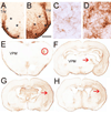
Figure 4. Immediate astrocytic response to neonatal ION damage all along the trigeminal pathway
A, B, Comparison of GFAP labeling in the denervated and unaffected PrVs from the same brain following unilateral neonatal ION damage. Asterisks indicate the borders of the ION-recipient zone of the PrV. C, D, Higher magnification views from the control and denervated PrV. Scale bar = 250 um for A, B, and 20 µm for C, D. E. Section through the thalamus showing conspicuous increase in GFAP immunostaining in the ION input recipient zone (red circle) of the VPM contralateral to the ION damaged side. F. A distinct GFAP immunopositivity is detectable in the contralateral VPM even at low magnification views of the brain sections. G, H, two representative sections showing large areas of the parietal cortex, including the barrel cortex, with high levels of GFAP immunoreactivity. These astrocytic responses occur rather fast after neonatal ION damage. The mechanisms underlying this transregional signaling, astrocytic response, and the consequences are largely unknown.

References

    1. Adams JC. Thrombospondins: multifunctional regulators of cell interactions. Annu. Rev. Cell Dev. Biol. 2001;17:25–51. - PubMed
    1. Alcalá-Galiano A, Arribas-García IJ, Martín-Pérez MA, Romance A, Montalvo-Moreno JJ, Juncos JM. Pediatric facial fractures: children are not just small adults. Radiographics. 2008;28:441–461. - PubMed
    1. Aldskogius H, Arvidsso J. Nerve cell degeneration and death in the trigeminal ganglion of the adult rat following peripheral nerve transection. J. Neurocytol. 1978;7:229–250. - PubMed
    1. Aldskogius H, Arvidsson J, Grant G. The reaction of primary sensory neurons to peripheral nerve injury with particular emphasis on transganglionic changes. Brain Res. 1985;357:27–46. - PubMed
    1. Allen NJ. Role of glia in developmental synapse formation. Curr. Opin. Neurobiol. 2013;23:1027–1033. - PubMed

Publication types