. 2015 May 9:15:381.
doi: 10.1186/s12885-015-1360-4.

Significance of KRAS/PAK1/Crk pathway in non-small cell lung cancer oncogenesis

Affiliations

Significance of KRAS/PAK1/Crk pathway in non-small cell lung cancer oncogenesis

Fariborz Mortazavi et al. BMC Cancer. .

Abstract

Background: Key effector(s) of mutated KRAS in lung cancer progression and metastasis are unknown. Here we investigated the role of PAK1/Crk axis in transduction of the oncogenic KRAS signal in non-small cell lung cancer (NSCLC).

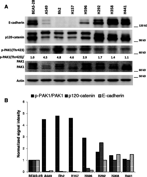
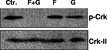
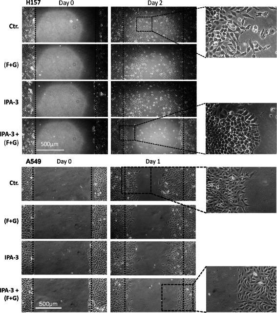
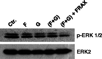
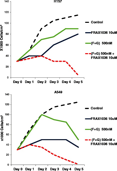
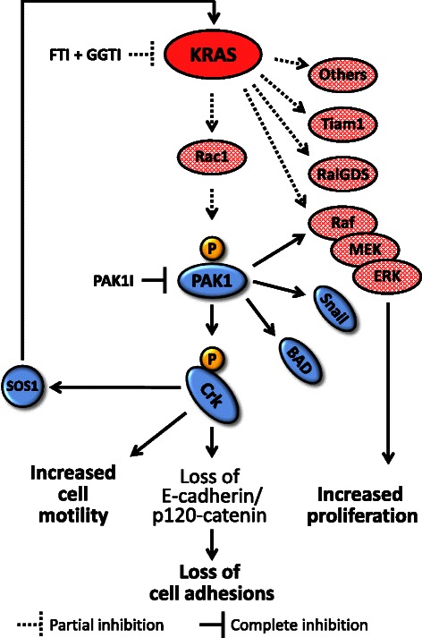
Methods: We used NSCLC clinical specimens to examine the correlation among KRAS mutations (codon 12, 13 and 61); PAK1/Crk axis activation [p-PAK1(Thr423), p-Crk(Ser41)]; and adhesion molecules expression by immunohistochemistry. For assessing the role of proto-oncogene c-Crk as a KRAS effector, we inhibited KRAS in NSCLC cells by a combination of farnesyltransferase inhibitor (FTI) and geranylgeranyltransferase inhibitor (GGTI) and measured p-Crk-II(Ser41) by western blotting. Finally, we disrupted the signaling network downstream of KRAS by blocking KRAS/PAK1/Crk axis with PAK1 inhibitors (i.e., IPA-3, FRAX597 or FRAX1036) along with partial inhibition of all other KRAS effectors by prenylation inhibitors (FTI + GGTI) and examined the motility, morphology and proliferation of the NSCLC cells.

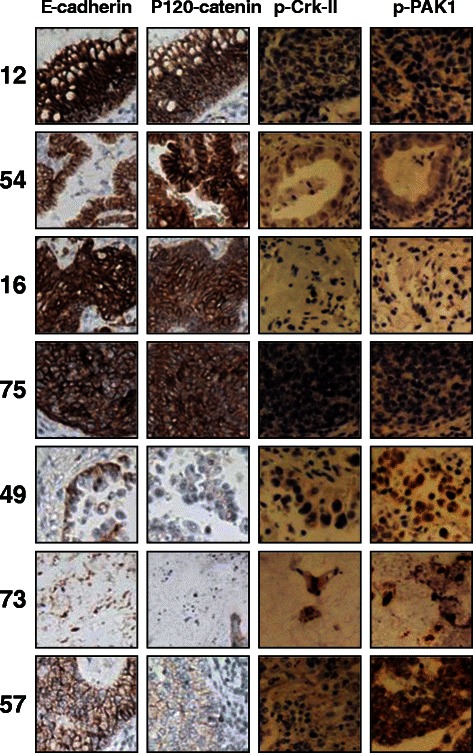
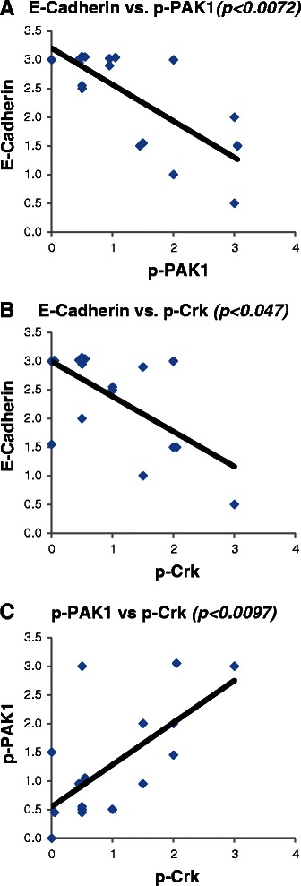
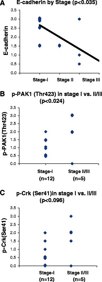
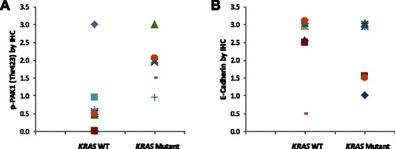
Results: Immunohistochemical analysis demonstrated an inverse correlation between PAK1/Crk phosphorylation and E-cadherin/p120-catenin expression. Furthermore, KRAS mutant tumors expressed higher p-PAK1(Thr423) compared to KRAS wild type. KRAS prenylation inhibition by (FTI + GGTI) completely dephosphorylated proto-oncogene c-Crk on Serine 41 while Crk phosphorylation did not change by individual prenylation inhibitors or diluent. Combination of PAK1 inhibition and partial inhibition of all other KRAS effectors by (FTI + GGTI) dramatically altered morphology, motility and proliferation of H157 and A549 cells.

Conclusions: Our data provide evidence that proto-oncogene c-Crk is operative downstream of KRAS in NSCLC. Previously we demonstrated that Crk receives oncogenic signals from PAK1. These data in conjunction with the work of others that have specified the role of PAK1 in transduction of KRAS signal bring forward the importance of KRAS/PAK1/Crk axis as a prominent pathway in the oncogenesis of KRAS mutant lung cancer.


Figures

Figure 1
Figure 1
PAK1 activation and Crk phosphorylation are correlated with loss of E-cadherin and p120-catenin in NSCLC specimens. Photomicrographs demonstrating immunohistochemical staining of NSCLC clinical specimens. Samples 12, 54, 16 and 75 harbor high E-cadherin/p120-catenin while expressing no detectable level of p-PAK1(Thr423) and p-Crk-II(Ser41). On the other hand, samples 49, 73 and 57 with detectable p-PAK1(Thr423) and p-Crk-II(Ser41) show very low levels of E-cadherin/p120-catenin.
Figure 2
Figure 2
PAK1 activation and Crk phosphorylation are positively correlated while establish a negative correlation with E-cadherin in NSCLC. Dot plots demonstrating the correlation between A-E-cadherin and p-PAK1(Thr423); B-E-cadherin and p-Crk-II(Ser41); C-p-PAK1(Thr423) and p-Crk-II(Ser41) expression by immunohistochemistry in 17 NSCLC clinical specimens. The average intensity of protein expression across the slide is quantified in a scale from 0-3+. The correlation between the expressions of each two marker was examined by utilizing Spearman Rank Correlation statistical test.
Figure 3
Figure 3
PAK1 activation is inversely correlated with E-cadherin and p120-catenin expression in NSCLC cells. A-Western blots demonstrating E-cadherin, p120-catenin, PAK1 and p-PAK1(Thr423) expression in a panel of NSCLC cells as well as immortalized normal human respiratory epithelial cells (BEAS-2B). B-Bar chart demonstrating signal intensity of p-PAK1/PAK1, p120-catenin and E-cadherin normalized to the value of BEAS-2B cells.
Figure 4
Figure 4
PAK1 activation is correlated with tumor stage at presentation. A-Dot plot demonstrating the expression of E-cadherin in the examined tumors in relation to the surgical stage of each tumor. The correlation between variables were examined by Spearman Rank Correlation analysis. B- Dot plot demonstrating the expression of p-PAK1(Thr423) in stage I and stage II/III tumors. The mean between groups was compared by student T-test. C- Dot plot demonstrating the expression of p-Crk-II(Ser41) in stage I and stage II/III tumors. The mean between groups was compared by student T-test.
Figure 5
Figure 5
KRAS mutant NSCLC specimens express higher activated PAK1 compared to KRAS wild type samples. A-Dot plot demonstrating immunohistochemical expression of p-PAK1(Thr423) in KRAS mutant and KRAS wild type NSCLC clinical specimens. p-PAK1(Thr423) is detectable in all KRAS mutant specimens while most KRAS wild type samples express lower p-PAK1(Thr423). B- Dot plot demonstrating immunohistochemical expression of E-cadherin in KRAS mutant vs. KRAS wild type NSCLC clinical specimens.
Figure 6
Figure 6
KRAS inhibition dephosphorylates Crk-II on serine 41. Western blots demonstrating loss of Crk-II phosphorylation on serine 41 in Rh2 NSCLC cells following 24 hour exposure of cells to combination of (F) farnesyltransferase inhibitor (BMS-225975 at 2 μM) and (G) geranylgeranyltransferase inhibitor (P61A6 at 2 μM).
Figure 7
Figure 7
Addition of PAK1 inhibitor to KRAS prenylation inhibitors dramatically alters cell morphology and motility. Photomicrographs of standard wound healing assays in H157 and A549 cells (KRAS mutant). Cells were exposed to (i) farnesyltransferase inhibitor (BMS-225975 at 2 μM) and geranylgeranyltransferase inhibitor (P61A6 at 2 μM) [F + G]; (ii) PAK1 inhibitor (IPA-3 at 5 μM); (iii) combination of the three inhibitors or (iv) the inhibitor vehicles for 24-48 hours.
Figure 8
Figure 8
Addition of PAK1 inhibitor to KRAS prenylation inhibitors dephosphorylated ERK. Western blots demonstrate ERK dephosphorylation following 24 hour exposure of Rh2 cells to (F) farnesyltransferase inhibitor (BMS-225975 at 2 μM), (G) geranylgeranyltransferase inhibitor (P61A6 at 2 μM), combination of the two (F + G), or (F + G) along with PAK1 inhibitor (FRAX597 at 40 μM).
Figure 9
Figure 9
Addition of PAK1 inhibitor to KRAS prenylation inhibitors alters the proliferation of NSCLC cells. Line charts demonstrating mean cell count of H157 and A549 cells upon exposure to prenylation inhibitors [FTI (BMS-225975) and GGTI (P61A6) at 500 nM each], PAK1 inhibitor (FRAX1036 at 10 μM) or combination in comparison to cells exposed to inhibitors’ diluent (DMSO).
Figure 10
Figure 10
Cooperative disruption of signaling network downstream of mutated KRAS. Schematic view of KRAS/PAK1/Crk signaling pathway is demonstrated. Partial inhibition of KRAS signaling output by tolerable dose of prenylation inhibitors (i.e., FTI + GGTI) in addition to inhibition of a prominent KRAS effector (i.e., PAK1) result in dramatic anti-tumor effects. FTI: farnesyltransferase inhibitor, GGTI: geranylgeranyltransferase inhibitor, PAK1I: p21 associated kinase 1 inhibitor.

Similar articles

Cited by

References

    1. Chow HY, Jubb AM, Koch JN, Jaffer ZM, Stepanova D, Campbell DA, et al. p21-Activated kinase 1 is required for efficient tumor formation and progression in a Ras-mediated skin cancer model. Cancer Res. 2012;72:5966–75. doi: 10.1158/0008-5472.CAN-12-2246. - DOI - PMC - PubMed
    1. McDaniel AS, Allen JD, Park SJ, Jaffer ZM, Michels EG, Burgin SJ, et al. Pak1 regulates multiple c-Kit mediated Ras-MAPK gain-in-function phenotypes in Nf1+/- mast cells. Blood. 2008;112:4646–54. doi: 10.1182/blood-2008-04-155085. - DOI - PMC - PubMed
    1. Singhal R, Kandel ES. The response to PAK1 inhibitor IPA3 distinguishes between cancer cells with mutations in BRAF and Ras oncogenes. Oncotarget. 2012;3:700–8. doi: 10.18632/oncotarget.587. - DOI - PMC - PubMed
    1. Tabusa H, Brooks T, Massey AJ. Knockdown of PAK4 or PAK1 inhibits the proliferation of mutant KRAS colon cancer cells independently of RAF/MEK/ERK and PI3K/AKT signaling. Mol Cancer Res. 2013;11:109–21. doi: 10.1158/1541-7786.MCR-12-0466. - DOI - PubMed
    1. Rettig M, Trinidad K, Pezeshkpour G, Frost P, Sharma S, Moatamed F, et al. PAK1 kinase promotes cell motility and invasiveness through CRK-II serine phosphorylation in non-small cell lung cancer cells. PLoS One. 2012;7:e42012. doi: 10.1371/journal.pone.0042012. - DOI - PMC - PubMed

Publication types

MeSH terms