. 2015 Aug;213(2):188.e1-188.e11.
doi: 10.1016/j.ajog.2015.05.001. Epub 2015 May 5.

Evaluating maternal recovery from labor and delivery: bone and levator ani injuries

Affiliations

Evaluating maternal recovery from labor and delivery: bone and levator ani injuries

Janis M Miller et al. Am J Obstet Gynecol. 2015 Aug.

Abstract

Objective: We sought to describe occurrence, recovery, and consequences of musculoskeletal (MSK) injuries in women at risk for childbirth-related pelvic floor injury at first vaginal birth.

Study design: Evaluating Maternal Recovery from Labor and Delivery is a longitudinal cohort design study of women recruited early postbirth and followed over time. We report here on 68 women who had birth-related risk factors for levator ani (LA) muscle injury, including long second stage, anal tears, and/or older maternal age, and who were evaluated by MSK magnetic resonance imaging at both 7 weeks and 8 months' postpartum. We categorized magnitude of injury by extent of bone marrow edema, pubic bone fracture, LA muscle edema, and LA muscle tear. We also measured the force of LA muscle contraction, urethral pressure, pelvic organ prolapse, and incontinence.

Results: In this higher-risk sample, 66% (39/59) had pubic bone marrow edema, 29% (17/59) had subcortical fracture, 90% (53/59) had LA muscle edema, and 41% (28/68) had low-grade or greater LA tear 7 weeks' postpartum. The magnitude of LA muscle tear did not substantially change by 8 months' postpartum (P = .86), but LA muscle edema and bone injuries showed total or near total resolution (P < .05). The magnitude of unresolved MSK injuries correlated with magnitude of reduced LA muscle force and posterior vaginal wall descent (P < .05) but not with urethral pressure, volume of demonstrable stress incontinence, or self-report of incontinence severity (P > .05).

Conclusion: Pubic bone edema and subcortical fracture and LA muscle injury are common when studied in women with certain risk factors. The bony abnormalities resolve, but levator tear does not, and is associated with levator weakness and posterior-vaginal wall descent.

Keywords: levator ani; magnetic resonance imaging; musculoskeletal injuries; pelvic floor; vaginal birth.


Conflict of interest statement

Conflict of Interest Disclosure: The authors report no conflict of interest.

Figures

Figure 1
Figure 1. 31 year-old with high-grade bone marrow edema and bilateral LA tears, right greater than left. By convention, images are presented in anatomic position and axial images as if viewed from the feet. 1A
Initial MRI with axial STIR sequence that demonstrates the right pubic bone marrow edema (horizontal arrow). STIR sequence images, such as this one, always appear coarse because of the poor spatial resolution inherent in the acquisition of these sequences. Although most clinicians will find these images less pleasing to study than the more familiar anatomic images, the STIR sequences have high sensitivity to sites of edema (edema as bright white contrast) compared with anatomic images. Vertical arrows indicate the bilateral LA tears (better seen in 1B). 1B. Initial MRI with axial proton density sequence showing bilateral LA tears. Vertical arrows are pointing to the region where thick LA muscle should be demonstrated between the vagina and the internal obturator muscle, but essentially, the muscle is missing for this person. Proton density sequences such as this one will always appear with better spatial resolution compared with STIR sequence, but the proton density sequences do not reveal the bone marrow edema that was seen in the STIR sequence of 1A. 1C. Follow-up MRI with axial proton density fat saturation sequence that demonstrates almost complete resolution of bone marrow edema. Image sequences of proton density with fat saturation appear less coarse than STIR sequences (in the 1A image) but still allow for better visualization of bone marrow edema than proton density-weighted image without fat suppression, shown in 1D. 1D. Follow-up MRI with axial proton density without fat saturation sequence showing bilateral LA tears (vertical arrows) unchanged. Arrowheads point to the inner margin of muscle site.
Figure 2
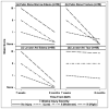
Figure 2. Mean MRI scores at two evaluation times, grouped by 7-weeks postpartum score
Subjects were grouped by magnitude level at 7-weeks postpartum; magnitude levels were ordered from 0 (none) to 3 (intense/severe). Each group starts at its group magnitude level because all women within a group have the same magnitude level at 7-weeks postpartum. Panels (a), (c), and (d) show improvement over time as the mean scores drop 0 (none). Magnitude level in panel (b) shows little improvement in LA tear, indicating toward grossly stable muscle tear to 8-months postpartum. Edema in the levator muscle or in the pubic bone marrow shows a pattern of resolving over time.
Figure 3
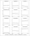
Figure 3. Mean values of clinical and questionnaire findings at 7-weeks postpartum and 8-months postpartum grouped by LA tear severity at 7-weeks postpartum
Each panel shows trends over time for variables relating to incontinence (standing stress test [PTT]; Antonakos, Sandvik, and Wexner), Kegel strength (resting and rep average measured by one-billed speculum), and POP-Q measures. Most measures do not show meaningful differences for different levels of LA tear severity, except in the case of the Kegel rep averages (p=0.006) and posterior vaginal wall descent (p=0.005).

Comment in

References

    1. Lien KC, Mooney B, DeLancey JO, Ashton-Miller JA. Levator ani muscle stretch induced by simulated vaginal birth. Obstet Gynecol. 2004;103:31–40. - PMC - PubMed
    1. DelGrande F, Santini F, Herzka D, Aro M, Dean C, Gold G, Carrino J. Fat-suppression techniques for 3-T MR imaging of the musculoskeletal system. Radiographics. 2014;34:217–233. - PMC - PubMed
    1. Nattiv A, Kennedy G, Barrack M, Abdelkerim A, Goolsby M, Arends J, Seeger L. Correlation of MRI grading of bone stress injuries with clinical risk factors and return to play: a 5-year prospective study in collegiate track and field athletes. Am J Sports Med. 2013;41:1930–1941. - PMC - PubMed
    1. Datir A, Saini A, Connell A, Saifuddin A. Stress-related bone injuries with emphasis on MRI. Clin Radiol. 2007;62:828–836. - PubMed
    1. Plewes D, Kuscharczyk W. Physics of MRI, a primer. J Magn Reson Imaging. 2012;35:1038–1054. - PubMed

Publication types

MeSH terms