

Microarchitectural and mechanical characterization of the sickle bone
- PMID: 25957113
- PMCID: PMC4442736
- DOI: 10.1016/j.jmbbm.2015.04.019
Microarchitectural and mechanical characterization of the sickle bone
Abstract
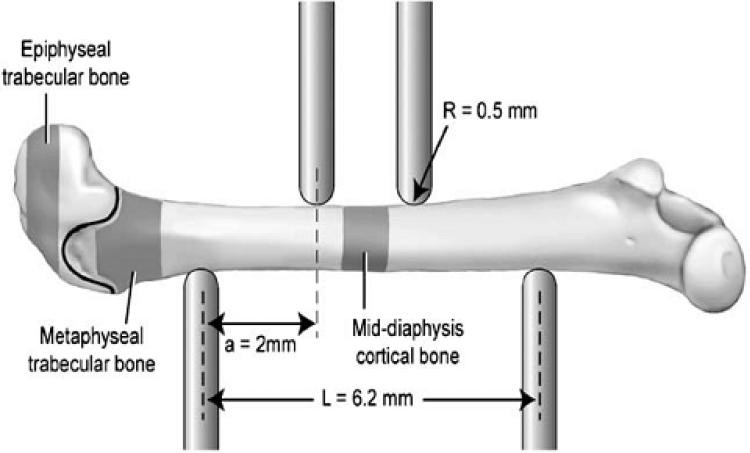
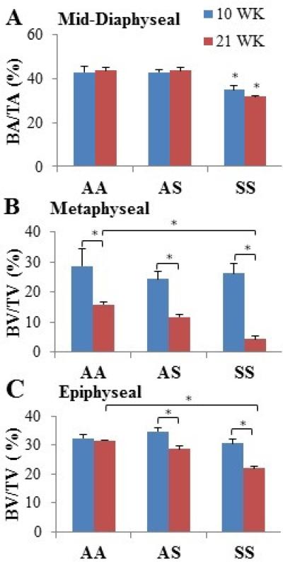
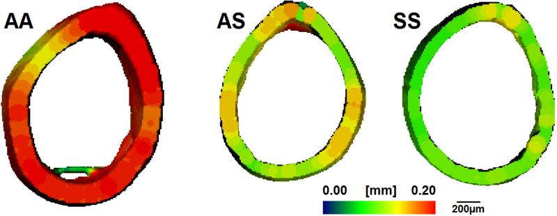
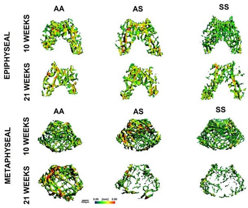
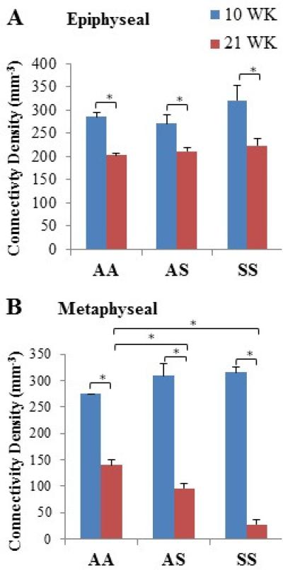
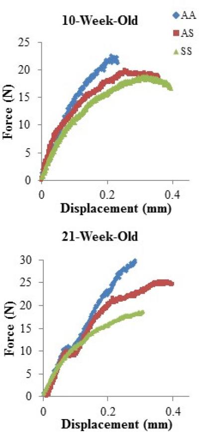
Individuals with sickle cell disease often experience acute and chronic bone pain due to occlusive events within the tissue vasculature that result in ischemia, necrosis, and organ degeneration. Macroscopically, sickle bone is identified in clinical radiographs by its reduced mineral density, widening of the marrow cavity, and thinning of the cortical bone due to the elevated erythroid hyperplasia accompanying the disease. However, the microstructural architecture of sickle bone and its role in mechanical functionality is largely unknown. This study utilized micro-CT and biomechanical testing to determine the relationship between the bone morphology, tissue mineral density, and trabecular and cortical microarchitecture of 10- and 21-week-old femurs from transgenic sickle male mice and littermates with sickle trait, as well as a wild-type control. While bone tissue mineral density did not vary among the genotypes at either age, variation in bone microstructure were observed. At 10 weeks, healthy and trait mice exhibited similar morphology within the cortical and trabecular bone, while sickle mice exhibited highly connected trabeculae. Within older femurs, sickle and trait specimens displayed significantly fewer trabeculae, and the remaining trabeculae had a more deteriorated geometry based on the structure model index. Thinning of the cortical region in sickle femurs contributed to the displayed flexibility with a significantly lower elastic modulus than the controls at both 10- and 21-weeks old. Wild-type and trait femurs generally demonstrated similar mechanical properties; however, trait femurs had a significantly higher modulus than sickle and wild-type control at 21-weeks. Overall, these data indicate that the progressive damage to the microvasculature caused by sickle cell disease, results in deleterious structural changes in the bone tissue׳s microarchitecture and mechanics.
Keywords: Biomechanics; Bone; Micro-CT; Microarchitecture; Sickle cell disease.
Copyright © 2015 Elsevier Ltd. All rights reserved.
Figures
References
-
- Aaron JE, et al. The microanatomy of trabecular bone loss in normal aging men and women. Clinical Orthopedics. 1987;215:260–271. - PubMed
-
- Adewoye A, et al. Sickle cell bone disease: Response to vitamin D and calcium. American Journal of Hematology. 2008;83:271–274. - PubMed
-
- Aguilar C, et al. Bone and joint disease in sickle cell disease. Hematology/Oncology Clinics of North America. 2005;(19):929–941. - PubMed
-
- Almedia A, Roberts I. Bone involvement in sickle cell disease. British Journal of Haematology. 2005;129:482–490. - PubMed
-
- Arlet J, et al. Relationship between vitamin D deficiency and bone fragility in sickle cell disease: A cohort study adults. Bone. 2013;52:206–211. - PubMed
Publication types
MeSH terms
Grants and funding
LinkOut - more resources
Full Text Sources
Other Literature Sources
Medical
