. 2015 Jun;25(6):836-44.
doi: 10.1101/gr.188581.114. Epub 2015 May 8.

Genomic redistribution of GR monomers and dimers mediates transcriptional response to exogenous glucocorticoid in vivo

Affiliations

Genomic redistribution of GR monomers and dimers mediates transcriptional response to exogenous glucocorticoid in vivo

Hee-Woong Lim et al. Genome Res. 2015 Jun.

Abstract

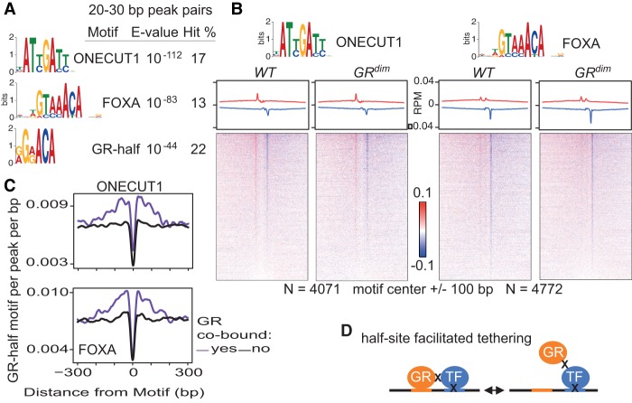
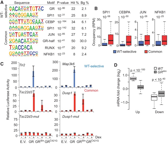
Glucocorticoids (GCs) are commonly prescribed drugs, but their anti-inflammatory benefits are mitigated by metabolic side effects. Their transcriptional effects, including tissue-specific gene activation and repression, are mediated by the glucocorticoid receptor (GR), which is known to bind as a homodimer to a palindromic DNA sequence. Using ChIP-exo in mouse liver under endogenous corticosterone exposure, we report here that monomeric GR interaction with a half-site motif is more prevalent than homodimer binding. Monomers colocalize with lineage-determining transcription factors in both liver and primary macrophages, and the GR half-site motif drives transcription, suggesting that monomeric binding is fundamental to GR's tissue-specific functions. In response to exogenous GC in vivo, GR dimers assemble on chromatin near ligand-activated genes, concomitant with monomer evacuation of sites near repressed genes. Thus, pharmacological GCs mediate gene expression by favoring GR homodimer occupancy at classic palindromic sites at the expense of monomeric binding. The findings have important implications for improving therapies that target GR.


Figures

Figure 1.
Figure 1.
GR, but not GRdim, occupies the canonical palindromic motif as a dimer in liver. (A) Top-ranked de novo motifs from HOMER for the GR cistromes from WT and GRdim mice. See Supplemental Material for a comprehensive list of motifs. (B) Scatter plots comparing sequence tags from 14,940 GR ChIP-seq peaks with at least two reads per million (RPM) in any condition in livers isolated from WT and GRdim mice killed at either 6 a.m. (top) or 6 p.m. (bottom). Blue and red highlight WT-selective and common sites, respectively. (C) GR binding upstream of the tyrosine aminotransferase (Tat) gene. The 5′ ends of forward- and reverse-stranded sequence tags are indicated in red and blue, respectively, for the ChIP-exo tracks. Tracks are RPM normalized. (D) Distance distribution for opposite-stranded peaks with at least 0.2 RPM from GR ChIP-exo in liver isolated at 6 a.m. from WT and GRdim mice is shown for WT-selective sites. The number of peak pairs and prominent peak distances are indicated. Schematic of opposite-stranded peaks is shown at top. (E) GR ChIP-exo for WT-selective sites in liver isolated at 6 a.m. MEME top-ranked de novo sequence with a hit count of at least 5% is shown at the top. See Supplemental Material for a full list of motifs. Average profiles (middle) and density heatmaps (bottom) of the raw sequence tags are shown for both mouse models. Red and blue indicate the 5′ ends of the forward- and reverse-stranded tags, respectively.
Figure 2.
Figure 2.
GR and GRdim occupy the liver genome at the half-site motif. (A) The distance distribution for opposite-stranded peaks with at least 0.2 RPM from GR ChIP-exo in liver isolated at 6 a.m. is shown for sites commonly bound in WT and GRdim mice, with the number of peak pairs and prominent peak distances indicated. (B) MEME de novo sequences from 6 a.m. common site peak pairs separated by 5–15 bp and with a hit count of at least 5%. See Supplemental Material for a comprehensive list of motifs. (C) GR ChIP-exo at 6 a.m. common sites, with average profiles and density heatmaps for the half-site motif shown for both mouse models. Red and blue indicate the 5′ ends of the forward- and reverse-stranded tags, respectively. (D) Distribution of the GR-half motif relative to the center of GR ChIP-seq peaks for the common sites subdivided into quartiles. (E) Plot of GR-binding strength relative to the number of half-site motifs present in ChIP-seq peaks from common sites.
Figure 3.
Figure 3.
GR and GRdim occupy the liver genome at tethered sites. (A) MEME de novo sequences from 6 a.m. common site peak pairs separated by 20–30 bp and with a hit count of at least 5%. See Supplemental Material for a comprehensive list of motifs. (B) GR ChIP-exo at 6 a.m. common sites, with average profiles and density heatmaps for the ONECUT1 (left) and FOXA (right) motifs shown for both mouse models. GR common sites cobound by ONECUT1 or FOXA2 were interrogated. Red and blue indicate the 5′ ends of the forward- and reverse-stranded tags, respectively. (C) Distribution of the GR half-site motif relative to neighboring motifs at common sites cobound by ONECUT1 (top) or FOXA2 (bottom). Results for ONECUT1 and FOXA2 liver sites without GR are shown for comparison. (D) Half-site-facilitated tethering. The GR half-site motif is represented in orange. X indicates formaldehyde crosslinking between proteins or protein–DNA. Two formaldehyde crosslinking events between GR and a DNA-crosslinked TF are necessary to detect sites where GR appears bound to noncanonical motifs.
Figure 4.
Figure 4.
Monomeric GR colocalizes with lineage-determining TFs in liver. (A) HOMER de novo motif analyses for the dimeric and monomeric GR binding sites from liver ChIP-seq. The four top-ranked lineage TF motifs are shown relative to the top-ranked GR sequence. See Supplemental Material for a comprehensive list of motifs. (B) Box plots interrogating the co-occupancy of liver TFs at GR binding sites. (C) Distribution of dimer and monomer GR binding sites relative to colocalized HNF4A, CEBPB, ONECUT1, and/or FOXA2. All TF combinations were examined.
Figure 5.
Figure 5.
GRdim partitions the GR cistrome in primary macrophages. (A) Top-ranked de novo motifs from HOMER for the WT-selective and common GR binding sites from ChIP-seq in primary macrophages. Comprehensive motif results are reported in the Supplemental Material. (B) Box plots interrogating the co-occupancy of macrophage TFs at GR binding sites. (C) Regions named after the nearest gene that include WT-selective and common sites were assayed by a luciferase reporter in the absence (−) or presence (+) of dexamethasone (Dex) and cotransfection of empty vector (E.V.), GR, GRdim, or the DNA-binding mutant GRC421G. (D) Response of WT-regulated genes (up or down) from LPS- and dex-treated macrophages in GRdim-derived macrophages under the same treatment.
Figure 6.
Figure 6.
Glucocorticoids (GCs) redistribute GR from monomeric to dimeric sites at regulated genes. (A) Density heatmap of ChIP-seq reads from GR peaks at 6 a.m. with at least 2 RPM in any condition. Prednisolone (pred) treatment of WT and GRdim mice spanned 24 h, and regulated sites in WT mice have at least a twofold difference compared with untreated controls. Top-ranked GO terms for the gained and lost sites are indicated on the left. MEME top-ranked de novo sequences are indicated on the right. See Supplemental Material for a comprehensive list of motifs. (B) GR ChIP-exo at gained sites in WT mice with or without Pred treatment. Average profiles (top) and density heatmaps (bottom) show the 5′ ends of the forward-stranded (red) and reverse-stranded (blue) sequence tags. (C) Box plot comparing CEBPB occupancy at sites with increased GR binding (gained) after Pred treatment. (D) Scatter plot comparing sequence tags from GR ChIP-seq in WT liver with and without Pred treatment, with sites containing up- or down-regulated RNAPII occupancy in response to Pred highlighted in red or blue, respectively. (E) Fraction of Pred-regulated genes with a transcription start site within 100 kb of a gained or lost GR binding site (top) or a non-Pred-regulated dimeric or monomeric GR binding site (bottom).

References

    1. Adams M, Meijer OC, Wang J, Bhargava A, Pearce D. 2003. Homodimerization of the glucocorticoid receptor is not essential for response element binding: activation of the phenylethanolamine N-methyltransferase gene by dimerization-defective mutants. Mol Endocrinol 17: 2583–2592. - PubMed
    1. Barish GD, Downes M, Alaynick WA, Yu RT, Ocampo CB, Bookout AL, Mangelsdorf DJ, Evans RM. 2005. A Nuclear Receptor Atlas: macrophage activation. Mol Endocrinol 19: 2466–2477. - PubMed
    1. Baschant U, Frappart L, Rauchhaus U, Bruns L, Reichardt HM, Kamradt T, Bräuer R, Tuckermann JP. 2011. Glucocorticoid therapy of antigen-induced arthritis depends on the dimerized glucocorticoid receptor in T cells. Proc Natl Acad Sci 108: 19317–19322. - PMC - PubMed
    1. Baschant U, Lane NE, Tuckermann J. 2012. The multiple facets of glucocorticoid action in rheumatoid arthritis. Nat Rev Rheumatol 8: 645–655. - PubMed
    1. Beck IME, Vanden Berghe W, Vermeulen L, Yamamoto KR, Haegeman G, De Bosscher K. 2009. Crosstalk in inflammation: the interplay of glucocorticoid receptor-based mechanisms and kinases and phosphatases. Endocr Rev 30: 830–882. - PMC - PubMed

Publication types

MeSH terms

Associated data