. 2015 May 9:11:26.
doi: 10.1186/s12990-015-0024-3.

Sodium channel Nav1.7 in vascular myocytes, endothelium, and innervating axons in human skin

Affiliations

Sodium channel Nav1.7 in vascular myocytes, endothelium, and innervating axons in human skin

Frank L Rice et al. Mol Pain. .

Abstract

Background: The skin is a morphologically complex organ that serves multiple complementary functions, including an important role in thermoregulation, which is mediated by a rich vasculature that is innervated by sympathetic and sensory endings. Two autosomal dominant disorders characterized by episodes of severe pain, inherited erythromelalgia (IEM) and paroxysmal extreme pain disorder (PEPD) have been directly linked to mutations that enhance the function of sodium channel Nav1.7. Pain attacks are accompanied by reddening of the skin in both disorders. Nav1.7 is known to be expressed at relatively high levels within both dorsal root ganglion (DRG) and sympathetic ganglion neurons, and mutations that enhance the activity of Nav1.7 have been shown to have profound effects on the excitability of both cell-types, suggesting that dysfunction of sympathetic and/or sensory fibers, which release vasoactive peptides at skin vasculature, may contribute to skin reddening in IEM and PEPD.

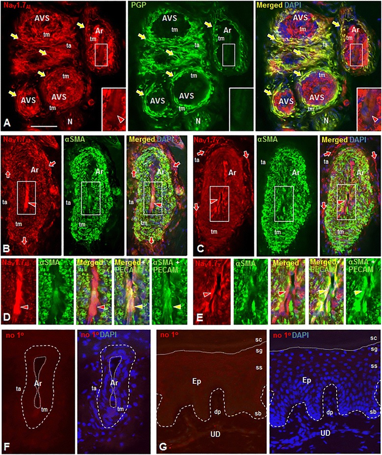
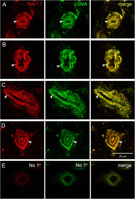
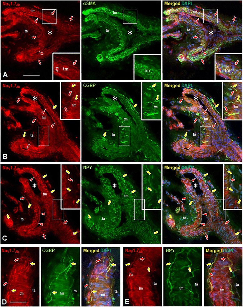
Results: In the present study, we demonstrate that smooth muscle cells of cutaneous arterioles and arteriole-venule shunts (AVS) in the skin express sodium channel Nav1.7. Moreover, Nav1.7 is expressed by endothelial cells lining the arterioles and AVS and by sensory and sympathetic fibers innervating these vascular elements.

Conclusions: These observations suggest that the activity of mutant Nav1.7 channels in smooth muscle cells of skin vasculature and innervating sensory and sympathetic fibers contribute to the skin reddening and/or pain in IEM and PEPD.


Figures

Figure 1
Figure 1
Nav1.7 immunolabeling (IL) of arterioles (Ar), arteriole-venule shunts (AVS) and associated innervation in normal human plantar glabrous skin with Alomone (A, B) or Yale (C) Nav1.7 antibodies (red). Co-labeling of innervation (arrows) as marked with anti-PGP 9.5 (PGP, green, A) or smooth muscle cells in tunica media (tm) as marked with anti α-smooth muscle actin antibody (αSMA, green, B,C). Nuclei are DAPI-labeled (blue). Left images (each panel) show only red fluorescence, middle images green; right images show triple-label combinations. Large white rectangles are 2X-enlargements of small rectangles. A-C. Nav1.7-IL is expressed on endothelial cells of tunica intima (red arrowheads) and tm smooth muscle cells as confirmed by double-labeling with anti-αSMA (B, C). Nav1.7-IL is expressed on virtually all vascular innervation (arrows) in tunica adventitia (ta) as confirmed by anti-PGP 9.5 double-labeling (A, yellow arrows). N=nerve. D-E. Nav1.7-IL on arteriole endothelial cells shown as 2X-enlargements of areas indicated by white rectangles in B,C. First images (each panel) show Nav1.7-IL on smooth muscle cells in tm and endothelial cells (red arrowheads). The second images show α-SMA co-labeling of only the smooth muscle cells of tm (green). The third images show merge of first and second images with DAPI (blue). Sections re-labeled with anti-PECAM (green) to show co-labeling with Nav1.7 on endothelial cells (yellow arrowheads, fourth and fifth images). F-G. Background Cy3 fluorescence is limited with no primary antibody in arteriole deep in dermis (F), epidermis (Ep) and upper-dermis (UD) (G). In F, broken line shows tm perimeter with dotted line around arteriole lumen. In G, broken line indicates basement membrane of epidermis and dotted line indicates boundary of dead and live superficial keratinocyte layers (stratum corneum, sc and stratum granulosum, sg, respectively). Stratum spinosum, ss; stratum basalis, sb; dermal papilla (dp). Scale bars=150μm (A); 100μm (B ,C, F, G); 50μm in D,E.
Figure 2
Figure 2
Nav1.7 expression in smooth muscle cells of deep dermis arterioles within skin from lateral malleolus of three healthy subjects. Smooth muscle cells (arrowheads) of the arteriole tunica media exhibit robust Nav1.7 (red) immunolabeling (antibody Nav1.7Y), which is co-localized with alpha smooth muscle actin (green). Skin samples from 3 healthy subjects (Subject 1: A; Subject 2: B,C; Subject 3: D) display similar patterns of Nav1.7 labeling in the smooth muscle cells of the dermal arterioles. Co-localization of Nav1.7 and alpha smooth muscle actin is yellow in the merged panels. E. Sections incubated without primary antibodies followed by secondary antibodies displayed background levels of immunofluorescence in skin vasculature.
Figure 3
Figure 3
Digital fluorescence images of Nav1.7 immunolabeling (IL) of arterioles (Ar), arteriole-venule shunts (AVS) and associated innervation in normal human glabrous skin biopsies from the plantar foot (A,C,D) and palmar hand (B). All sections are labeled with an Alomone (A,C) or Yale (B,D) rabbit anti-rat Nav1.7 antibody revealed by a donkey anti-rabbit Cy3-conjugated secondary antibody (red fluorescence). Secondary antibodies conjugated to Alexa 488 (green fluorescence) were used to assess co-labeling for peptidergic sensory innervation revealed with a sheep anti-human CGRP antibody (A,B) or noradrenergic sympathetic innervation revealed with a sheep anti-human NPY antibody (C,D). Cell nuclei are labeled with DAPI (blue fluorescence). The left images in each panel show only the red fluorescence, the middle images only the green, and the right images the triple label combinations. Areas outlined in large white rectangles are 2X enlargement of the areas in the small rectangles. A-D. Nav1.7-IL is expressed on the endothelial cells of the tunica intima (red arrowheads) and on smooth muscle cells of the tunica media (tm). A,B. Peptidergic sensory innervation co-expresses Nav1.7-IL and CGRP-IL (yellow straight arrows). Other innervation labeled only with Nav1.7 (red curved arrows) is likely the noradrenergic sympathetic innervation that expresses NPY-IL as shown in C and D. C,D. Noradrenergic sympathetic innervation co-expresses Nav1.7-IL and NPY-IL (yellow curved arrows). Other innervation labeled with only Nav1.7 (red straight arrows) is likely the peptidergic sensory innervation that expresses CGRP-IL as shown in A and B. Scale Bar = 150 μm in A and B, 100 μm in C and D.
Figure 4
Figure 4
Nav1.7 immunolabeling (IL) of arterioles and associated innervation in normal human palmar glabrous skin biopsies, in alternating sections cut parallel to and through lumen (*) of branched arteriole (A-C) and parallel to arteriole, skimming the interface between tunica media (tm) and tunica adventitia (ta) (D,E). All sections are labeled with Abcam anti-human Nav1.7 antibody (red). Secondary antibodies conjugated to Alexa 488 (green) were used to assess co-labeling for: smooth muscle cells revealed with mouse anti-α−smooth muscle actin antibody (αSMA, A); peptidergic sensory innervation revealed with sheep anti-CGRP antibody (yellow straight arrows, B,D); and noradrenergic sympathetic innervation revealed with sheep anti-NPY antibody (yellow curved arrows, C,E): Nuclei are labeled with DAPI (blue). Left images in each panel show only the red fluorescence, middle images only green, and right images the triple-label combinations. Areas outlined in large white rectangles (A-C) are 2X enlargements of areas in small rectangles. A-E. A. Nav1.7-IL is expressed on endothelial cells of tunica intima (red arrowheads) and smooth muscle cells of tm as confirmed by double-labeling with anti-αSMA. Nav1.7-IL is expressed on innervation (arrows) in ta, near and at the border with tm. B, D. Peptidergic sensory innervation co-expresses Nav1.7-IL and CGRP-IL (yellow straight arrows). Other innervation labeled only with Nav1.7 (red curved arrows) is likely noradrenergic sympathetic innervation that expresses NPY-IL (C,E). C,E. Noradrenergic sympathetic innervation co-expresses Nav1.7-IL and NPY-IL (yellow curved arrows). Other innervation labeled with only Nav1.7 (red straight arrows) is likely peptidergic sensory innervation that expresses CGRP-IL as shown in B and D. Scale bar = 100 μm in A-C, 50 μm in D and E.
Figure 5
Figure 5
Digital fluorescence images of Nav1.7 (red) and PGP 9.5 (green) immunolabeling (IL) in the epidermis (Ep) and upper dermis (UD) biopsies of normal human palmar glabrous skin (A, Abcam anti-Nav1.7) and normal human plantar glabrous skin (B, Alomone anti-Nav1.7). Stratum corneum, sc; stratum granulosum, sg; stratum spinosum (ss); stratum basalis (sb), dermal papilla (dp). Straight arrows indicate epidermal sensory endings, curved arrows indicate small nerves and individual axons or endings in the upper dermis. The areas enclosed in the large rectangles are 2X enlargements of those in the smaller rectangles. Of all the innervation revealed by anti-PGP 9.5, only some express Nav1.7-IL (yellow straight and curved arrows) whereas other only express PGP 9.5-IL (green straight and curved arrows). Aβ-fiber innervation of a Meissner corpuscle (MC) has little if any Nav1.7-IL. Kertinocytes especially in stratum granulosum label for Nav1.7 (arrowheads) which has a more membranous distribution with the Alomone anti-Nav1.7 antibody, but more diffuse labeling with the Abcam anti-Nav1.7 antibody. Scale bar = 100 μm.

References

    1. Rice F, Albrecht P. Cutaneous Mechanisms of Tactile Perception: Morphological and Chemical Organization of the Innervation to the Skin. In: Basbaum A, Kaneko A, Shepard G, Westheimer G, editors. The Senses: A comprehensive Reference, vol 6, Somatosensation. San Diego: Academic Press; 2008. pp. 1–32.
    1. Albrecht PJ, Hou Q, Argoff CE, Storey JR, Wymer JP, Rice FL. Excessive peptidergic sensory innervation of cutaneous arteriole-venule shunts (AVS) in the palmar glabrous skin of fibromyalgia patients: implications for widespread deep tissue pain and fatigue. Pain Med. 2013;14(6):895–915. - PubMed
    1. Burton AC. The range and variability of the blood flow in the human fingers and the vasomotor regulation of body temperature. Am J Physiol. 1939;127:437–53.
    1. Johnson JM, Kellogg DL., Jr Thermoregulatory and thermal control in the human cutaneous circulation. Front Biosci (Schol Ed) 2010;2:825–53. - PubMed
    1. Kellogg DL., Jr In vivo mechanisms of cutaneous vasodilation and vasoconstriction in humans during thermoregulatory challenges. J Appl Physiol. 2006;100(5):1709–18. - PubMed

Publication types

Substances