

Review
doi: 10.1101/cshperspect.a020487.
Schwann Cells: Development and Role in Nerve Repair
Affiliations
- PMID: 25957303
- PMCID: PMC4484967
- DOI: 10.1101/cshperspect.a020487
Item in Clipboard
Review
Schwann Cells: Development and Role in Nerve Repair
Cold Spring Harb Perspect Biol.
.
Abstract
Schwann cells develop from the neural crest in a well-defined sequence of events. This involves the formation of the Schwann cell precursor and immature Schwann cells, followed by the generation of the myelin and nonmyelin (Remak) cells of mature nerves. This review describes the signals that control the embryonic phase of this process and the organogenesis of peripheral nerves. We also discuss the phenotypic plasticity retained by mature Schwann cells, and explain why this unusual feature is central to the striking regenerative potential of the peripheral nervous system (PNS).
Copyright © 2015 Cold Spring Harbor Laboratory Press; all rights reserved.
Figures
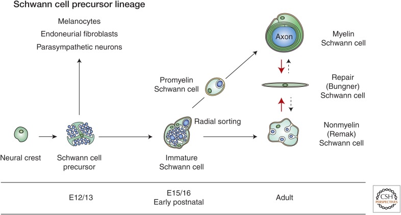
Main transitions in the Schwann cell precursor (SCP) lineage. The diagram shows both developmental and injury-induced transitions. Black uninterrupted arrows, normal development; red arrows, the Schwann cell injury response; stippled arrows, postrepair reformation of myelin and Remak cells. Embryonic dates (E) refer to mouse development. (Modified from Jessen and Mirsky 2012; reprinted, with permission and with contribution from Y. Poitelon and L. Feltri.)

Diagram showing the architecture and main cellular components of an adult peripheral nerve. The main cellular structures within the nerve and the connective tissue compartments and the perineurium that protects them are indicated. This nerve contains one fascicle; larger nerves consist of several fascicles embedded in a common epineurium. The perineurium shown here, as a single cell layer, is most often multilayered. The drawing does not show the basal lamina that surrounds individual Schwann cell/axon units, blood vessels, and perineurial cells.

The phenotype of key stages in embryonic Schwann cell development. Each stage involves characteristic relationships with surrounding tissues and distinctive signaling properties (indicated in the panels immediately below the lineage drawing). Also shown are some of the molecular markers of the lineage. They fall into three groups: (1) markers that show no significant change between the three stages; (2) markers that are up-regulated during development (some of these are up-regulated at the crest to Schwann cell precursor transition; another group is up-regulated at the Schwann cell precursor to immature Schwann cell transition); (3) markers that are down-regulated at the Schwann cell precursor to immature Schwann cell transition. Sch, Schwann cell. (Modified from Jessen and Mirsky 2005; reprinted, with permission. See the original reference for detailed references to the molecules shown.)

Schwann cell precursors (SCP) and immature Schwann cells (iSch) in embryonic nerves. (Upper panel) Transverse section of E14 rat sciatic nerve. Schwann cell precursors are embedded among the axons (downward large arrow) and at the surface of the nerve (upward large arrow). A dividing Schwann cell precursor is also seen (small arrow). Connective tissue (turquoise) is not found inside the nerve. (Lower panel) Transverse section of E18 rat sciatic nerve. One or a few immature Schwann cells together surround several axons, forming compact groups or families (asterisk). A dividing Schwann cell is seen (double arrows). Connective tissue (turquoise) containing blood vessels (large arrow) is present throughout the nerve surrounding the families. Bracket indicates the developing perineurium. (From Jessen and Mirsky 2005; adapted, with permission, from the authors.)

Key tissue components of regenerating nerves. (a) Regeneration unit, and (b) fibroblasts and immune cells providing essential signals to the Schwann cells of the regeneration units. In the distal stump, denervated Schwann cells convert to a repair supportive phenotype. These repair (Bungner) Schwann cells form regeneration tracks (Bungner bands; dark blue cells) that guide regenerating axons back to their targets and provide essential trophic support for injured neurons. After crush injury, axons are severed but the Schwann cell basal lamina tubes and connective sheaths remain continuous between the proximal and distal stump. This allows axons to reach the distal stump within their original basal lamina tubes. After nerve cut, the length of the bridge depends on the extent of the injury and the nature of any subsequent surgical intervention.
References
-
- Allodi I, Udina E, Navarro X. 2012. Specificity of peripheral nerve regeneration: Interactions at the axon level. Prog Neurobiol 98: 16–37. - PubMed
-
- Armstrong SJ, Wiberg M, Terenghi G, Kingham PJ. 2007. ECM molecules mediate both Schwann cell proliferation and activation to enhance neurite outgrowth. Tissue Eng 13: 2863–2870. - PubMed
-
- Atanasoski S, Shumas S, Dickson C, Scherer SS, Suter U. 2001. Differential cyclin D1 requirements of proliferating Schwann cells during development and after injury. Mol Cell Neurosci 18: 581–592. - PubMed
Publication types
MeSH terms
Grants and funding
LinkOut - more resources
Full Text Sources
Other Literature Sources