

Building the Microtubule Cytoskeleton Piece by Piece
- PMID: 25957410
- PMCID: PMC4498055
- DOI: 10.1074/jbc.R115.638452
Building the Microtubule Cytoskeleton Piece by Piece
Abstract
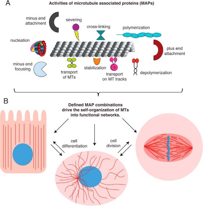
The microtubule (MT) cytoskeleton gives cells their shape, organizes the cellular interior, and segregates chromosomes. These functions rely on the precise arrangement of MTs, which is achieved by the coordinated action of MT-associated proteins (MAPs). We highlight the first and most important examples of how different MAP activities are combined in vitro to create an ensemble function that exceeds the simple addition of their individual activities, and how the Xenopus laevis egg extract system has been utilized as a powerful intermediate between cellular and purified systems to uncover the design principles of self-organized MT networks in the cell.
Keywords: cell division; cytoskeleton; microtubule; microtubule-associated protein (MAP); mitotic spindle.
© 2015 by The American Society for Biochemistry and Molecular Biology, Inc.
Figures
References
-
- Mitchison T., Kirschner M. (1984) Dynamic instability of microtubule growth. Nature 312, 237–242 - PubMed
-
- Desai A., Mitchison T. J. (1997) Microtubule polymerization dynamics. Annu. Rev. Cell Dev. Biol. 13, 83–117 - PubMed
-
- Nédélec F., Surrey T., Karsenti E. (2003) Self-organisation and forces in the microtubule cytoskeleton. Curr. Opin. Cell Biol. 15, 118–124 - PubMed
Publication types
MeSH terms
Substances
Grants and funding
LinkOut - more resources
Full Text Sources
