Review
. 2015 Jul 10;290(28):17163-72.
doi: 10.1074/jbc.R115.637447. Epub 2015 May 8.

Writing and Reading the Tubulin Code

Affiliations
Review

Writing and Reading the Tubulin Code

Ian Yu et al. J Biol Chem. .

Abstract

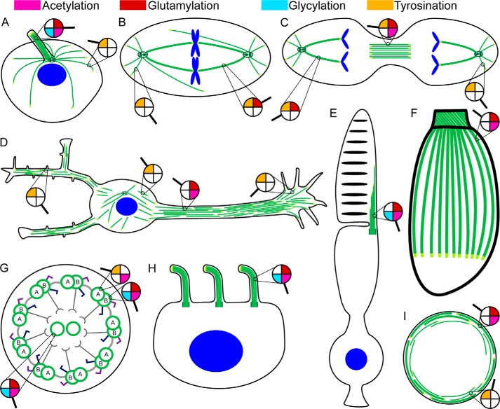
Microtubules give rise to intracellular structures with diverse morphologies and dynamics that are crucial for cell division, motility, and differentiation. They are decorated with abundant and chemically diverse posttranslational modifications that modulate their stability and interactions with cellular regulators. These modifications are important for the biogenesis and maintenance of complex microtubule arrays such as those found in spindles, cilia, neuronal processes, and platelets. Here we discuss the nature and subcellular distribution of these posttranslational marks whose patterns have been proposed to constitute a tubulin code that is interpreted by cellular effectors. We review the enzymes responsible for writing the tubulin code, explore their functional consequences, and identify outstanding challenges in deciphering the tubulin code.

Keywords: TTLL; cytoskeleton; microtubule; microtubule dynamics; microtubule motor; microtubule-associated protein (MAP); post-translational modification (PTM); tubulin; tubulin post-translational modifications; tubulin tyrosine ligase.


Figures

FIGURE 1.
FIGURE 1.
Microtubules form complex arrays of spatio-temporally regulated supra-structures. A, radial interphase array. B, mitotic spindle array. C, midbody array. D, neuron with complex, parallel, and tiled array in the axon and mixed polarity array in dendrites. E, photoreceptor cells with a connecting cilium between their inner and outer segments. Individual microtubules extend to varying depths of the outer segment. F, protozoans contain a unique membrane-embedded array of subpellicular microtubules and an additional apical cylindrical structure termed the conoid that consists of unique comma-shaped open polymers formed from nine laterally associated tubulin protofilaments. G, cross-sectional view of the nine-fold symmetric axonemal array. Light gray, nexin linkers; dark gray, radial spokes; dark blue, inner arm dyneins; purple, outer arm dyneins. H, cell with multiple motile cilia. I, marginal microtubule band in platelets; stable microtubules are coiled in the peripheral edges, whereas dynamic, tyrosinated microtubules actively polymerize/depolymerize. Microtubules are shown in green; microtubule plus-ends are in light green, and nuclei are in blue. The distribution of tubulin posttranslational modifications in the various microtubule arrays is indicated by a magnifying glass (pink, acetylation; red, glutamylation; cyan, glycylation; orange, tyrosination).
FIGURE 2.
FIGURE 2.
Posttranslational modifications map to both the body and the tails of the αβ-tubulin heterodimer. A, ribbon representation of the tubulin heterodimer (green, α-tubulin; blue, β-tubulin) with the disordered tubulin tails shown schematically using sequences for the α1A and βIVb tubulin isoforms. Sites of acetylation and polyamination are shown in stick representation (magenta and dark blue, respectively). The α-tubulin C-terminal tyrosine (orange) is subject to enzymatic removal/ligation (detyrosination/tyrosination cycle). Tail glutamates are subject to monoglutamylation and polyglutamylation (n denotes the number of glutamates in the elongated chain). Tail glutamates are also subject to monoglycylation and polyglycylation (m denotes the number of glycines in the elongated chain). B, zoomed-in view showing the acetylated β-tubulin Lys-252. C and D, view of the α-tubulin (C) and β-tubulin (D) longitudinal interfaces showing the position of mapped polyamination sites as dark blue sticks. α-Tubulin Lys-40 is shown in stick representation (magenta). Am, amination. Ac, acetylation.

References

    1. Mitchison T., Kirschner M. (1984) Microtubule assembly nucleated by isolated centrosomes. Nature 312, 232–237 - PubMed
    1. Gittes F., Mickey B., Nettleton J., Howard J. (1993) Flexural rigidity of microtubules and actin filaments measured from thermal fluctuations in shape. J. Cell Biol. 120, 923–934 - PMC - PubMed
    1. Janke C., Rogowski K., Wloga D., Regnard C., Kajava A. V., Strub J. M., Temurak N., van Dijk J., Boucher D., van Dorsselaer A., Suryavanshi S., Gaertig J., Eddé B. (2005) Tubulin polyglutamylase enzymes are members of the TTL domain protein family. Science 308, 1758–1762 - PubMed
    1. Rogowski K., Juge F., van Dijk J., Wloga D., Strub J. M., Levilliers N., Thomas D., Bré M. H., Van Dorsselaer A., Gaertig J., Janke C. (2009) Evolutionary divergence of enzymatic mechanisms for posttranslational polyglycylation. Cell 137, 1076–1087 - PubMed
    1. Rogowski K., van Dijk J., Magiera M. M., Bosc C., Deloulme J. C., Bosson A., Peris L., Gold N. D., Lacroix B., Bosch Grau M., Bec N., Larroque C., Desagher S., Holzer M., Andrieux A., Moutin M. J., Janke C. (2010) A family of protein-deglutamylating enzymes associated with neurodegeneration. Cell 143, 564–578 - PubMed

Publication types