Review
. 2015 Aug;149(2):302-17.e1.
doi: 10.1053/j.gastro.2015.04.053. Epub 2015 May 7.

Epidemiology, Diagnosis, and Management of Esophageal Adenocarcinoma

Affiliations
Review

Epidemiology, Diagnosis, and Management of Esophageal Adenocarcinoma

Joel H Rubenstein et al. Gastroenterology. 2015 Aug.

Abstract

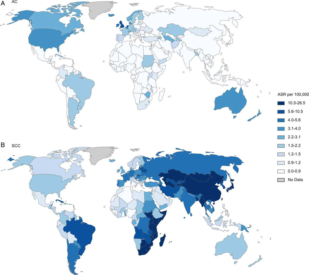
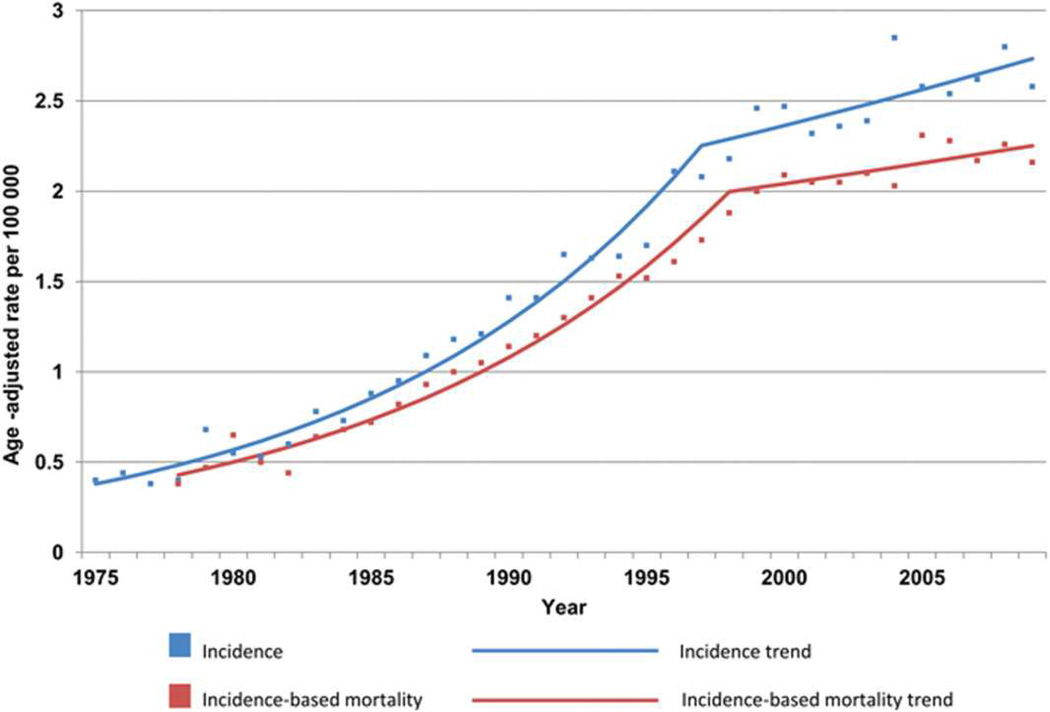
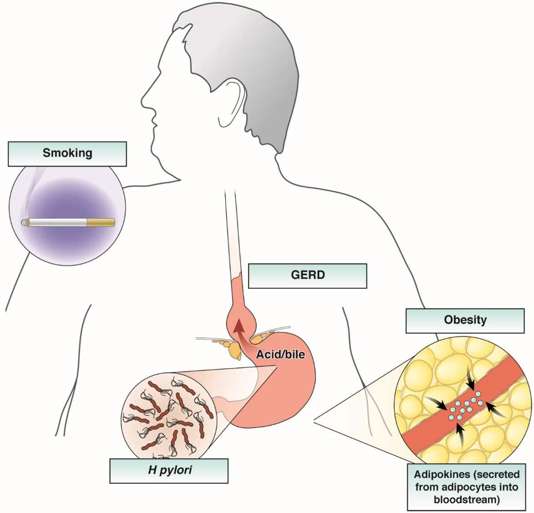
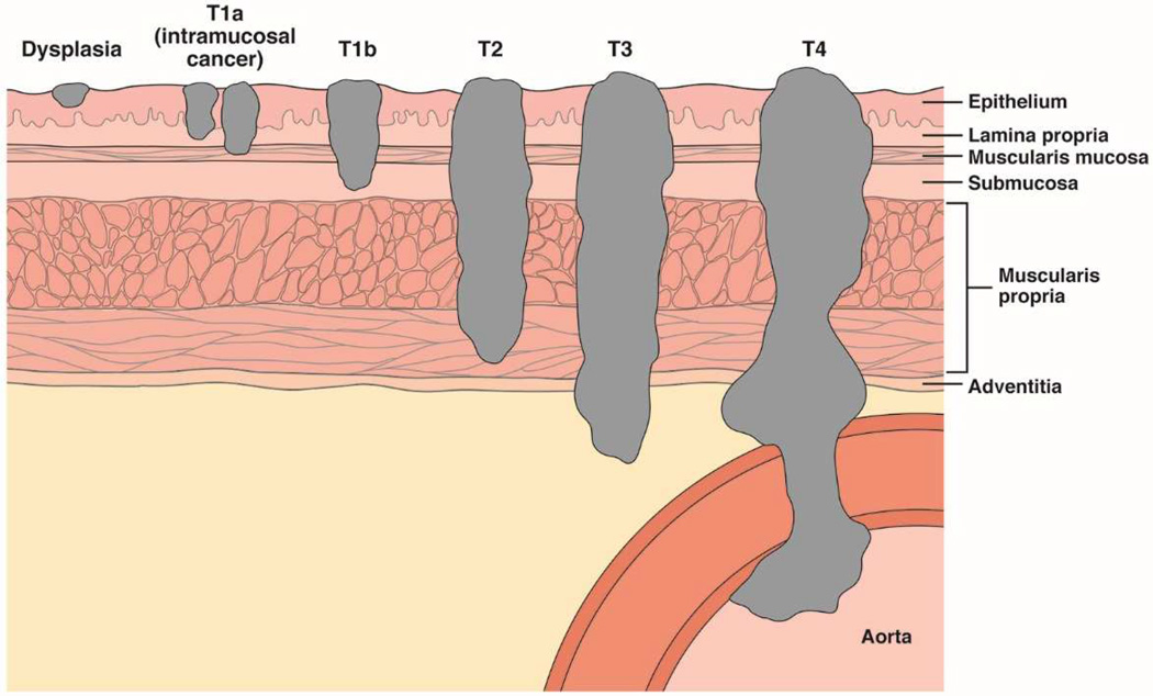
Esophageal adenocarcinoma (EAC) is rapidly increasing in incidence in Western cultures. Barrett's esophagus is the presumed precursor lesion for this cancer. Several other risk factors for this cancer have been described, including chronic heartburn, tobacco use, white race, and obesity. Despite these known associations, most patients with EAC present with symptoms of dysphagia from late-stage tumors; only a small number of patients are identified by screening and surveillance programs. Diagnostic analysis of EAC usually commences with upper endoscopy followed by cross-sectional imaging. Endoscopic ultrasonography is useful to assess the local extent of disease as well as the involvement of regional lymph nodes. T1a EAC may be treated endoscopically, and some patients with T1b disease may also benefit from endoscopic therapy. Locally advanced disease is generally managed with esophagectomy, often accompanied by neoadjuvant chemoradiotherapy or chemotherapy. The prognosis is based on tumor stage; patients with T1a tumors have an excellent prognosis, whereas few patients with advanced disease have long-term survival.

Keywords: Adenocarcinoma; Endoscopic Therapy; Esophageal Neoplasms; Risk Factors.


Figures

Figure 1
Figure 1. Global Differences in Incidence of Histological Subtypes of Esophageal Cancer
Age-standardized incidence rate (ASR) per 100,000 of (A) esophageal adenocarcinoma and (B) squamous cell carcinoma in men. AC, adenocarcinoma; SCC, squamous cell carcinoma. (Reproduced with permission from: Arnold M, Soerjomataram I, Ferlay J, et al. Global incidence of oesophageal cancer by histological subtype in 2012. Gut published online 10/15/2014 as doi:10.1136/gutjnl-2014-308124).
Figure 2
Figure 2. Trend in US Incidence of EAC
Graph shows Surveillance Epidemiology and End Results (SEER) cancer registry 9 esophageal adenocarcinoma incidence and incidence-based mortality, 1975 to 2009. From 1975 to 1997, EAC incidence increased at an annual percentage change (APC) of 8.4 (95% confidence interval [CI] = 7.7–9.1), whereas the APC was 1.6 (95% CI = 0.0–3.3) from 1997 to 2009. For incidence-based mortality, the APC was 8.0 from 1978 to 1998 (95% CI = 7.2–8.8) and 1.1 from 1998 to 2009 (95% CI = 0.7 to 2.9). All rates were age-adjusted to the 2000 Standard population using 19 age groups. (Reproduced with permission from: Hur C, Miller M, Kong CY, et al. Trends in Esophageal Adenocarcinoma Incidence and Mortality. Cancer 2013;119:1149–58).
Figure 3
Figure 3. Risk Factors for EAC
The primary risk factors for esophageal adenocarcinoma include male sex, advancing age, white race, GERD, obesity, and tobacco use. The effect of obesity is likely mediated both through a mechanical effect promoting GERD and a hormonal effect through alterations in circulating adipokines and other peptides, and appears to be a major risk factor for both BE and EAC. A deranged gastro-esophageal junction, noted above with a large hiatal hernia, allows free reflux of gastric contents. H pylori infection protects against EAC, and Barrett’s esophagus is its only known precursor.
Figure 4
Figure 4. Tumor Depth Staging for EAC
There are 4 main layers of the esophageal wall: mucosa, submucosa, muscularis propria, and adventitia. The mucosa is further divided into the epithelium, lamina propria, and muscularis mucosae. Dysplasia is confined to the epithelium. Intramucosal tumors (T1a) invade the lamina propria or muscularis mucosae. Tumors that invade the submucosa are classified T1b. T2 tumors invade the muscularis propria, T3 tumors invade the adventitia, and T4 tumors invade adjacent structures.
Figure 5
Figure 5. Endoscopic Management of Early EAC
Depth of invasion, as assessed by EMR or ESD, is the key to appropriate subsequent therapy. EMR, endoscopic mucosal resection; ESD, endoscopic submucosal dissection; LVI, lymphovascular invasion.

References

    1. Surveillance, Epidemiology, and End Results (SEER) Program ( www.seer.cancer.gov) SEER*Stat Database: Incidence - SEER 18 Regs Research Data + Hurricane Katrina Impacted Louisiana Cases, Nov 2013 Sub (2000–2011) <Katrina/Rita Population Adjustment> - Linked To County Attributes - Total U.S., 1969–2012 Counties.: National Cancer Institute, DCCPS, Surveillance Research Program. Cancer Statistics Branch. Released April 2014 (updated 5/7/2014), based on the November 2013 submission.
    1. Abrams JA, Sharaiha RZ, Gonsalves L, et al. Dating the rise of esophageal adenocarcinoma: analysis of Connecticut Tumor Registry data, 1940–2007. Cancer Epidemiology, Biomarkers & Prevention. 2011;20:183–186. - PMC - PubMed
    1. Trivers KF, Sabatino SA, Stewart SL, et al. Trends in esophageal cancer incidence by histology, United States, 1998–2003. International Journal of Cancer. 2008;123:1422–1428. - PubMed
    1. Devesa SS, Blot WJ, Fraumeni JF., Jr Changing patterns in the incidence of esophageal and gastric carcinoma in the United States. Cancer. 1998;83:2049–2053. - PubMed
    1. National Cancer Institute. Surveillance, Epidemiology and End Results (SEER) Program. [accessed January 9, 2015];Esophagus Table 8.22. www.seer.cancer.gov/csr/1975_2010/browse_csr.php.

Publication types

Supplementary concepts