. 2015 Jul:79:111-22.
doi: 10.1016/j.nbd.2015.04.011. Epub 2015 May 6.

Botulinum toxin in migraine: Role of transport in trigemino-somatic and trigemino-vascular afferents

Affiliations

Botulinum toxin in migraine: Role of transport in trigemino-somatic and trigemino-vascular afferents

Roshni Ramachandran et al. Neurobiol Dis. 2015 Jul.

Abstract

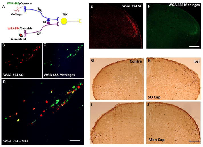
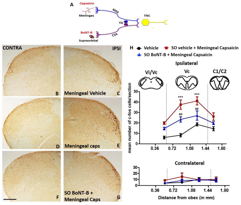
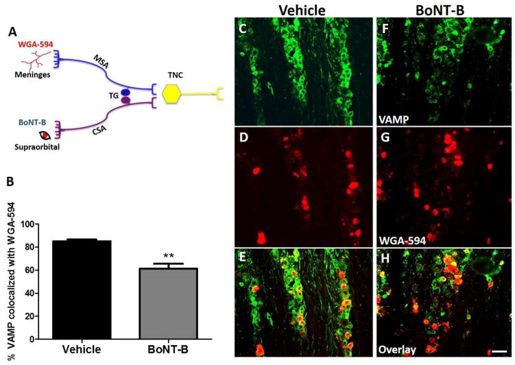
Migraine secondary to meningeal input is referred to extracranial regions innervated by somatic afferents that project to homologous regions in the trigeminal nucleus caudalis (TNC). Reported efficacy of extracranial botulinum toxin (BoNT) in treating migraine is surprising since a local extracranial effect of BoNT cannot account for its effect upon meningeal input. We hypothesize that intradermal BoNT acts through central transport in somatic afferents. Anesthetized C57Bl/6 mice (male) received unilateral supraorbital (SO) injections of BoNT-B (1.5 U/40 μl) or saline. 3 days later, mice received ipsilateral (ipsi)-SO capsaicin (20 μl of 0.5mM solution) or meningeal capsaicin (4 μl of 0.35 μM). Pre-treatment with ipsi-SO BoNT-B i) decreased nocicsponsive ipsilateral wiping behavior following ipsi-SO capsaicin; ii) produced cleavage of VAMP in the V1 region of ipsi-TG and in TG neurons showing WGA after SO injection; iii) reduced expression of c-fos in ipsi-TNC following ipsi-SO capsaicin; iv) reduced c-fos activation and NK-1 internalization in ipsi-TNC secondary to ipsi-meningeal capsaicin; and vi) SO WGA did not label dural afferents. We conclude that BoNT-B is taken up by peripheral afferents and transported to central terminals where it inhibits transmitter release resulting in decreased activation of second order neurons. Further, this study supports the hypothesis that SO BoNT exerts a trans-synaptic action on either the second order neuron (which receives convergent input from the meningeal afferent) or the terminal/TG of the converging meningeal afferent.

Keywords: Botulinum toxin; Migraine; Referred pain; Trans-synaptic; Transcytosis.


Conflict of interest statement

Conflict of Interest

We declare that there is no conflict of financial interest with regard to or manuscript.

Figures

Figure 1
Figure 1. Meningeal and supraorbital projections to TG and TNC
Schematic representation of experimental design where transganglionic tracer wheat germ agglutinin (WGA) tagged to fluorophores 594 or 488 and capsaicin was applied to the supraorbital (SO) and/or the meninges (dura mater) (A). Localization of WGA flurophores in the trigeminal ganglion (TG) (B–D). Note that the cutaneous sensory afferent neurons and meningeal sensory afferent neurons lie in close proximity to each other in the V1 region of TG. The nerve fibers in the ventrolateral region along lamina I and II of trigeminal nucleus caudalis (TNC) showed localization of WGA fluorophores (E, F). Localization of c-fos expression in TNC following SO (G, H) and meningeal capsaicin (I, J), N=4. Note both of the fluorophores stained nerve fibers localized in the same region as c-fos suggesting that the nerve fibers converge around the same second order neurons. CSA- cutaneous sensory afferents, MSA- meningeal sensory afferents, Men cap – meningeal capsaicin. Scale bar 100 μm.
Figure 2
Figure 2. Effects of supraorbital BoNT-B on ipsilateral supraorbital capsaicin induced pain behavior
Schematic representation of experimental design where the mice were treated with SO BoNT-B (1.5 U) or vehicle 3 days (3d) prior to ipsilateral SO capsaicin (20 μl of 0.5 mM) (A). Pain behavior measured by number of wipes following SO capsaicin injection (B), N=8. ***P<0.001.
Figure 3
Figure 3. Effects of supraorbital BoNT-B on VAMP expression in the trigeminal ganglion (TG)
Schematic representation of experimental design where the mice were treated with SO BoNT-B (1.5 U) or vehicle and sacrificed 3 days later (A). Longitudinal section (10 μm thickness) of TG showing V1 (ophthalmic), V2 (maxillary) and V3 (mandibular) region labelled with Neu N. Representative images of TG labelled with Neu N (red) and VAMP (green) in the V1 region following vehicle (C–E) and BoNT-B treatment (F–H). Histogram depicts the percentage of VAMP positive neurons in V1 region of TG. Note loss of VAMP is an indirect measure of VAMP cleavage by BoNT-B (I). Representative images of TG labelled with Neu N (red) and VAMP (green) in the V1 (J–L) and V3 (M–O) region of the same animal following BoNT-B treatment. Histogram depicts the percentage of VAMP positive neurons that was analyzed by the presence of VAMP (measure of VAMP cleavage by BoNT-B) in Neu N positive cells in the V1 and V3 (P) region following BoNT-B SO administration, N=4. **P<0.01 as compared to vehicle, *P<0.05 as compared to V3. TG- trigeminal ganglion, BoNT-B- botulinum toxin B, VAMP- vesicle associated membrane protein. Scale bar 75 μm.
Figure 4
Figure 4. Percentage of VAMP positive neurons co-localized with WGA-594 (transganglionic tracer injected in SO) in V1 (ophthalmic) region of trigeminal ganglion (TG)
Schematic representation of experimental design where the mice were supraorbitally injected with WGA-594, 1 day prior to ipsilateral SO BoNT-B (1.5 U) or vehicle (A). Histogram depicts the percentage of VAMP positive neurons that was colocalized with the WGA-594 positive neurons (reterogradely labelled from sensory cutaneous afferents (CSA)) in V1 region of TG. Loss of VAMP was the measure of VAMP cleavage (B). Representative images of TG labelled with VAMP (green) and WGA -594 (red) in the V1 (C–H) region, N=3. *P<0.05 as compared to vehicle. BoNT-B- botulinum toxin B, VAMP- vesicle associated membrane protein, V1- ophthalmic region, WGA- wheat germ agglutinin. Scale bar 75 μm.
Figure 5
Figure 5. Effects of supraorbital BoNT-B on c-fos expression evoked by ipsilateral supraorbital capsaicin in the trigeminal nucleus caudalis (TNC)
Schematic representation of experimental design where the mice were treated with SO BoNT-B (1.5 U) or vehicle 3 days (3d) prior to ipsilateral SO capsaicin (20 μl of 0.5 mM) (A). Representative images of ipsi and contra TNC labelled with c-fos positive cells in the Vc region treated with SO vehicle (B, C), SO capsaicin (D, E) and SO BoNT-B pretreatment followed by ipsi SO capsaicin (F, G). Graph represents the number of c-fos positive neurons in the ipsilateral and contralateral TNC rostrocaudally between 0.36 mm to 1.80 mm caudal to obex at 2 hrs after SO vehicle or capsaicin and following the pre-treatment with BoNT-B (1.5 U. 3 days earlier) (H). N=5–9. * P<0.05, ***P<0.001 as compared to SO vehicle. ###P<0.001 as compared to SO capsaicin. BoNT-B- botulinum toxin B. Scale bar 100 μm.
Figure 6
Figure 6. Effects of supraorbital BoNT-B on c-fos expression evoked by ipsilateral meningeal capsaicin in the trigeminal nucleus caudalis (TNC)
Schematic representation of experimental design where the mice were treated with SO BoNT-B (1.5 U) or vehicle 3 days (3d) prior to ipsilateral meningeal capsaicin (4 μl of 0.35 μM) (A). Representative images of ipsi and contra TNC labelled with c-fos positive cells in the ventrolateral region treated with meningeal vehicle (B, C), meningeal capsaicin (D, E) and SO BoNT-B pretreatment followed by ipsi meningeal capsaicin (F, G). Graph represents the number of c-fos positive neurons in the ipsilateral and contralateral TNC rostrocaudally between 0.36 mm to 1.80 mm caudal to obex at 2 hrs after meningeal vehicle or capsaicin and following the pre-treatment with BoNT-B (1.5 U, 3 days earlier) (H). N=6. ***P<0.001 as compared to meningeal vehicle. ##P<0.01 as compared to meningeal capsaicin. Scale bar 100 μm.
Figure 7
Figure 7. Effects of supraorbital BoNT-B on NK-1 receptor internalization in TNC induced by ipsilateral meningeal capsaicin
Schematic representation of experimental design where the mice were treated with SO BoNT-B (1.5 U) or vehicle 3 days (3d) prior to ipsilateral meningeal capsaicin (4 μl of 0.35 μM) (A). Plot represents percentage of NK-1 positive neurons that are internalized (marker for substance P release) at 15 min in TNC following meningeal capsaicin alone or group pretreated with SO BoNT-B, 3 days prior to ipsilateral meningeal capsaicin (B). Representative images of a neuron in ipsilateral ventrolateral TNC with internalized NK-1 receptor after meningeal capsaicin (C–H) and a non-internalized neuron in the TNC pretreated with SO BoNT-B prior to meningeal capsaicin (I–N). Note that represents neurons that are NK-1 receptor internalized and represents non-internalized NK-1 positive neurons. Note that pre-treatment with ipsilateral SO BoNT/B significantly decreased NK-1 internalization in these neurons (N=4). Scale bar C–E, I–K 100 μm and F–H, L–N 25 μm. ***P<0.001 as compared to meningeal capsaicin. BoNT-B- botulinum toxin B, NK-1 neurokinin 1 receptor, caps – capsaicin, SO- supraorbital, N=4.
Figure 8
Figure 8. Percentage of VAMP positive neurons co-localized with WGA-594 (applied in meninges (dura mater)) in V1 region of trigeminal ganglion (TG)
Schematic representation of experimental design where WGA-594 was applied in the dura mater, 1 day prior to ipsilateral SO BoNT-B (1.5 U) (A). Histogram depicts the percentage of VAMP positive neurons that was colocalized with the WGA-594 positive neurons (reterogradely labelled from meningeal sensory afferents (MSA)) in V1 region of TG. Note that loss of VAMP was the measure of VAMP cleavage (B). Representative images of TG labelled with VAMP (green) and WGA -594 (red) in the V1 (C–H) region, N=3. **P<0.01 as compared to vehicle. BoNT-B- botulinum toxin B, VAMP- vesicle associated membrane protein, V1- ophthalmic region, WGA- wheat germ agglutinin. Scale bar 75 μm.
Figure 9
Figure 9. Labelling of nerve fibers in dura mater following WGA-594 (transganglionic tracer) applied in SO and meninges
Representative image of nerve fibers close to MMA in the dura mater following SO WGA 594, tissue was co-stained with CGRP (A, B). Note that no dural fibers stained positively for WGA-594 (A), suggesting that meningeal collaterals do not project to the SO skin. Retrograde labelling of WGA-594 in the nerve fibers close to MMA was observed only after its application directly to the dura matter (C). This region is close to the site of WGA-594 applied in the meninges, N=3. Scale bar 50 μm.

Similar articles

Cited by

References

    1. Antonucci F, Rossi C, Gianfranceschi L, Rossetto O, Caleo M. Long-distance retrograde effects of botulinum neurotoxin A. J Neurosci. 2008;28(14):3689–3696. - PMC - PubMed
    1. Aoki KR. Evidence for antinociceptive activity of botulinum toxin type A in pain management. Headache. 2003;43(Suppl 1):S9–15. - PubMed
    1. Bach-Rojecky L, Lackovic Z. Antinociceptive effect of botulinum toxin type a in rat model of carrageenan and capsaicin induced pain. Croat Med J. 2005;46(2):201–208. - PubMed
    1. Bach-Rojecky L, Lackovic Z. Central origin of the antinociceptive action of botulinum toxin type A. Pharmacol Biochem Behav. 2009;94(2):234–238. - PubMed
    1. Bach-Rojecky L, Salkovic-Petrisic M, Lackovic Z. Botulinum toxin type A reduces pain supersensitivity in experimental diabetic neuropathy: bilateral effect after unilateral injection. Eur J Pharmacol. 2010;633(1–3):10–14. - PubMed

Publication types

MeSH terms