. 2015 May 19;11(7):1018-30.
doi: 10.1016/j.celrep.2015.04.031. Epub 2015 May 7.

Direct Activation of STING in the Tumor Microenvironment Leads to Potent and Systemic Tumor Regression and Immunity

Affiliations

Direct Activation of STING in the Tumor Microenvironment Leads to Potent and Systemic Tumor Regression and Immunity

Leticia Corrales et al. Cell Rep. .

Abstract

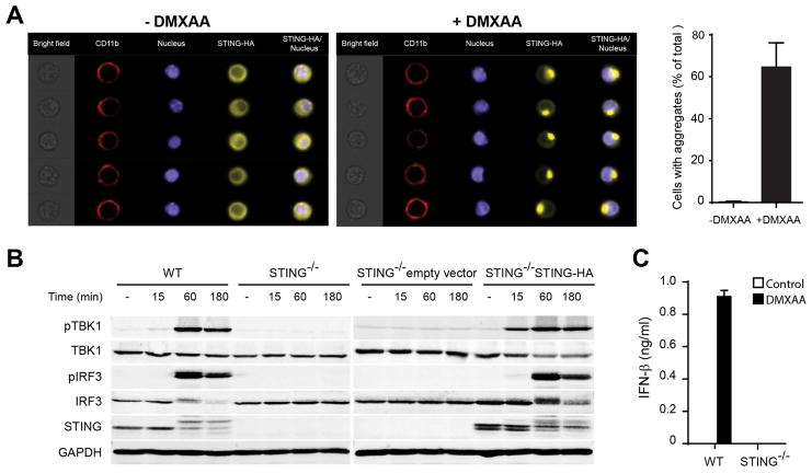
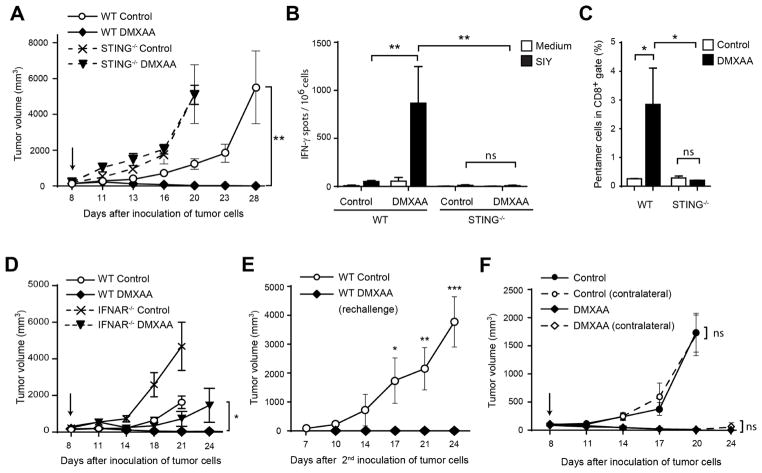
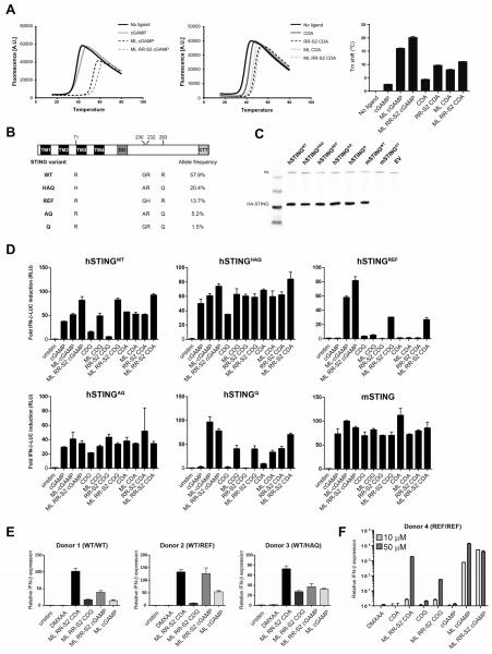
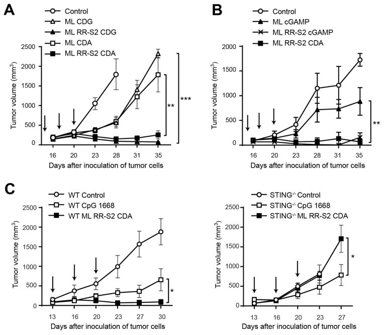
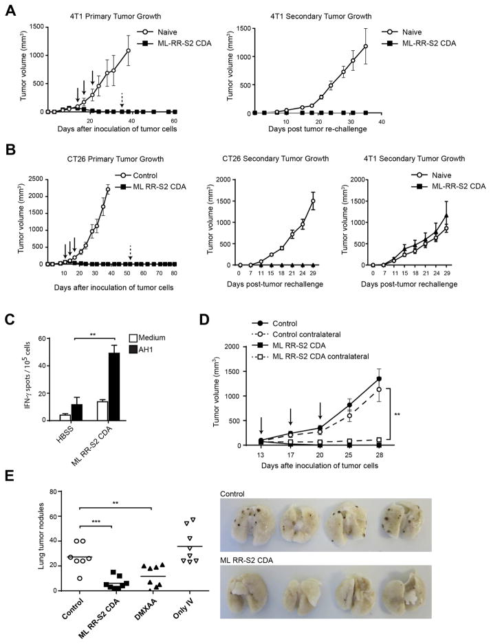
Spontaneous tumor-initiated T cell priming is dependent on IFN-β production by tumor-resident dendritic cells. On the basis of recent observations indicating that IFN-β expression was dependent upon activation of the host STING pathway, we hypothesized that direct engagement of STING through intratumoral (IT) administration of specific agonists would result in effective anti-tumor therapy. After proof-of-principle studies using the mouse STING agonist DMXAA showed a potent therapeutic effect, we generated synthetic cyclic dinucleotide (CDN) derivatives that activated all human STING alleles as well as murine STING. IT injection of STING agonists induced profound regression of established tumors in mice and generated substantial systemic immune responses capable of rejecting distant metastases and providing long-lived immunologic memory. Synthetic CDNs have high translational potential as a cancer therapeutic.


Conflict of interest statement

COMPETING FINANCIAL INTERESTS

L.H.G., S.M.M., D.B.K., K.E.S., G.E.K., E.L, T.B., J.J.L., K.M. and T.W.D are all paid employees of Aduro BioTech, hold stock in the company, and may be inventors on patent applications that apply to the CDN molecules described in the manuscript.

Figures

Figure 1
Figure 1. DMXAA activates the STING pathway and promotes the activation of APCs
(A) STING−/− mouse bone marrow-derived macrophages (BMM) transduced to express STING-HA tag were stimulated for 1 hour with 50 μg/ml DMXAA, stained with specific antibodies against HA-tag, CD11b and DAPI. Single cell images were acquired in the ImageStream and data were analyzed with the IDEAS software (Amnis, Millipore). The data in the graph represent average of percentage of cells with aggregates from three independent experiments. (B) WT or STING−/− BMM were stimulated with 50 μg/ml of DMXAA for the indicated time points. The amount of pTBK1, total TBK1, pIRF3, total IRF3, STING and GAPDH was measured by Western blot. (C) WT or STING−/− BMM were stimulated with 50 μg/ml of DMXAA for 12 hours. The amount of secreted IFN-β was measured by ELISA.
Figure 2
Figure 2. Rejection of tumors in response to DMXAA is STING-dependent
(A) WT or STING−/− C57BL/6 mice were inoculated with 106 B16.SIY cells in the left flank. When tumor volumes were 100–200 mm3 they received a single IT dose of 500 μg of DMXAA or saline. Tumor volume was measured at the indicated time points (n=5). (B–C) WT or STING−/− C57BL/6 mice (n=5) were treated as in (A) and 5 days later splenocytes were harvested and re-stimulated in vitro in the presence of culture medium or soluble SIY peptide for 16 hours. The frequency of tumor-specific IFN-γ–producing cells was assessed by ELISPOT (B), and the percentage of SIY-specific CD8+ T cells was assessed by staining splenocytes with antibodies against TCRβ, CD4, CD8 and SIY pentamer (C). Cells were acquired in the LSRII-Blue cytometer and analyzed with FlowJo software. Results are shown as mean ± s.e.m. * P < 0.05; ** P < 0.01. (D) WT or IFNAR−/− C57BL/6 mice were inoculated with 106 B16.SIY cells in the left flank (n=5). When tumor volumes were 100–200 mm3 they received a single IT dose of 500 μg of DMXAA or saline. Tumor volume was measured at the indicated time points. (E) WT mice that had rejected B16.SIY tumors were re-challenged with 106 B16.SIY in the contralateral flank. Naïve mice were used as controls. Tumor size was measured at the indicated time points. (F) WT mice were inoculated with 106 B16.SIY cells in the left and the right flanks (n=5). When tumor volumes were 100–200 mm3, 500 μg of DMXAA or saline was injected IT in the right flank only, and tumor volume was measured at the indicated time points. Data are representative of at least three independent experiments, or two independent experiments for the contralateral tumor model. Results are shown as mean tumor volume ± s.e.m. * P < 0.5; ** P < 0.01; *** P < 0.001. ns, not significant.
Figure 3
Figure 3. Modified CDNs potently activate STING and signal through all human STING allelic variants
(A) Purified human STING and murine STING binding to CDNs were analyzed by thermal shift assay. Temperature curves are the average from a representative experiment of three independent experiments performed in duplicate. Tm shift values are mean values + s.e.m. (B) Domain structure of hSTING is shown with the positions of the amino acid variations (bottom). The allelic frequencies of the hSTING isoforms shown on the left hand column were obtained from the 1000 Genome Project database as previously described (Yi et al., 2013). (C) HEK293T cells were stably transfected with the indicated STING alleles. Whole cell lysates from HEK293T cells stably expressing the indicated full length STING-HA proteins were analyzed by Western blot with anti-HA antibodies. (D) HEK293T cells expressing the indicated STING alleles were transfected with an IFN-β-luciferase reporter construct. After 24 hours, cells were stimulated for 6 hours with the indicated CDN compound (10 μM), and assessed for IFN-β-reporter activity. (E–F) Human PBMCs from donors with the indicated STING alleles were stimulated with 10 μM of the indicated CDN, or 100 μg/ml DMXAA (E), or human PBMCs from a donor homozygous for the reference variant (STINGREF/REF) was stimulated with 10 μM and 50 μM of the indicated CDN or 100 μg/ml DMXAA (F). After a 6 hour stimulation, fold induction of IFN-β was measured by q-RT-PCR and relative normalized expression was determined by comparison with untreated controls. Results are representative of at least two independent experiments.
Figure 4
Figure 4. Synthetic CDN modifications significantly improve anti-tumor efficacy in established B16 tumors
(A–B) WT C57BL/6 mice were inoculated with 5 × 10⁴ B16.F10 cells in the left flank (n=8). When tumor volumes were 100 mm3 they received three 25 μg IT doses of ML CDA, ML CDG, ML RR-S2 CDG, or ML RR-S2 CDA or HBSS as control (A); or either three 50 μg doses of the endogenous cGAS product ML cGAMP, ML RR-S2 CDA, ML RR-S2 cGAMP or HBSS as control (n=8) (B). (C) WT C57BL/6 mice or STING−/− mice were treated with three IT doses of CDN ML RR-S2 CDA (50 μg), murine type B CpG ODN 1668 (50 μg) or HBSS vehicle control. Treatments were administered on the days indicated by the arrows and tumor measurements were taken twice weekly. Data are representative of at least two independent experiments. Results are shown as mean tumor volume ± s.e.m. ** P < 0.01, *** P < 0.001. ns, not significant.
Figure 5
Figure 5. ML RR-S2 CDA promotes immune-mediated tumor rejection
(A–B) WT BALB/c mice were inoculated with 105 4T-1 breast cancer (A), or CT26 (B) colon carcinoma cells in the left flank. When tumor volumes were 100 mm3 they received three 50 μg doses IT of ML RR-S2 CDA, or HBSS vehicle control (left graph). Mice were re-implanted with 105 4T-1 (A and B); or CT26 (B) tumor cells on the opposite flank on day 55 post-initial tumor implantation. Naïve mice were used as controls (right graph) (n=8). (C) WT BALB/c mice were inoculated with 105 CT26 colon carcinoma cells in the left flank and treated on days 11, 14, and 18 with IT injections of ML RR-S2 CDA (25 μg each), or HBSS vehicle control (n= 4). 21 days post-implantation of CT26 tumors, PBMCs were stimulated with AH1 (gp70423–431) and assessed by IFN-γ ELISPOT assay. (D) WT BALB/c mice were implanted with 105 of CT26 tumor cells on both flanks. On the days indicated, mice were treated in one flank only with ML RR-S2 CDA (50 μg), or HBSS vehicle control (n=8). (E) WT C57BL/6 were inoculated with 5×10⁴ B16.F10 melanoma cells on the right flank at day 0, and implanted IV with 105 cells on day 7. Naïve mice were implanted with cells IV only as a control. Flank tumors were treated on the days indicated with ML RR-S2 CDA (50 μg), DMXAA (150 μg) or HBSS control (n=8). On day 28, lungs were harvested and lung tumor nodules counted. The histogram depicts total numbers of nodules in the ML RR-S2 CDA, DMXAA or HBSS control treated mice, compared to the untreated IV-only tumor implanted mice. The images depict the ML RR-S2 CDA and HBSS control treated mice. Data are representative of at least two independent experiments. Results are shown as mean ± s.e.m. ** P < 0.01, *** P < 0.001.

References

    1. Ablasser A, Goldeck M, Cavlar T, Deimling T, Witte G, Rohl I, Hopfner KP, Ludwig J, Hornung V. cGAS produces a 2′-5′-linked cyclic dinucleotide second messenger that activates STING. Nature. 2013;498:380–384. - PMC - PubMed
    1. Baguley BC, Ching LM. Immunomodulatory actions of xanthenone anticancer agents. BioDrugs: clinical immunotherapeutics, biopharmaceuticals and gene therapy. 1997;8:119–127. - PubMed
    1. Blank C, Brown I, Peterson AC, Spiotto M, Iwai Y, Honjo T, Gajewski TF. PD-L1/B7H-1 inhibits the effector phase of tumor rejection by T cell receptor (TCR) transgenic CD8+ T cells. Cancer Res. 2004;64:1140–1145. - PubMed
    1. Brody JD, Ai WZ, Czerwinski DK, Torchia JA, Levy M, Advani RH, Kim YH, Hoppe RT, Knox SJ, Shin LK, et al. In situ vaccination with a TLR9 agonist induces systemic lymphoma regression: a phase I/II study. J Clin Oncol. 2010;28:4324–4332. - PMC - PubMed
    1. Burdette DL, Monroe KM, Sotelo-Troha K, Iwig JS, Eckert B, Hyodo M, Hayakawa Y, Vance RE. STING is a direct innate immune sensor of cyclic di-GMP. Nature. 2011;478:515–518. - PMC - PubMed

Publication types

MeSH terms