. 2015 May 11:6:7097.
doi: 10.1038/ncomms8097.

Structural basis for dynamic mechanism of nitrate/nitrite antiport by NarK

Affiliations

Structural basis for dynamic mechanism of nitrate/nitrite antiport by NarK

Masahiro Fukuda et al. Nat Commun. .

Abstract

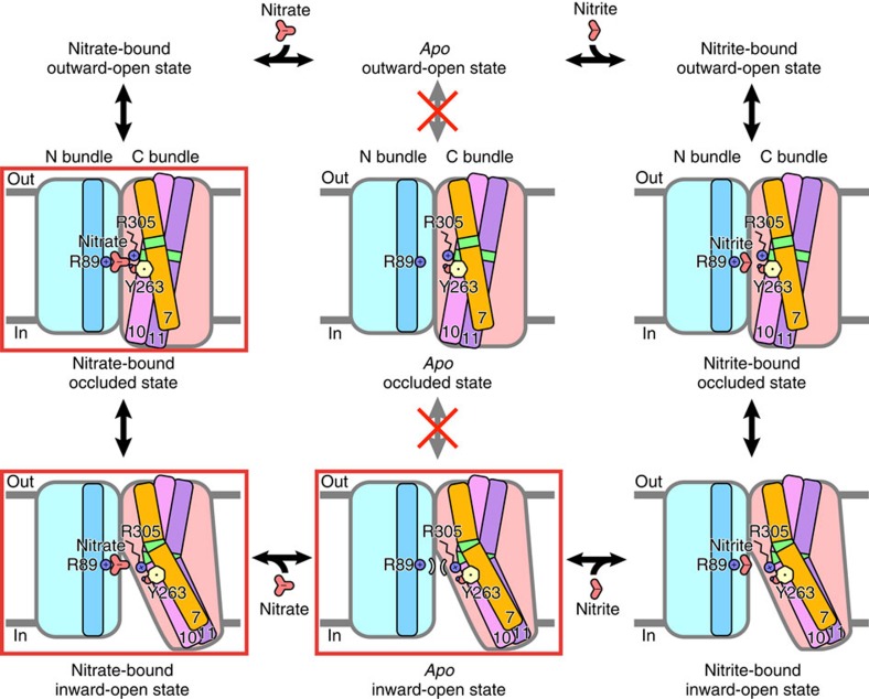
NarK belongs to the nitrate/nitrite porter (NNP) family in the major facilitator superfamily (MFS) and plays a central role in nitrate uptake across the membrane in diverse organisms, including archaea, bacteria, fungi and plants. Although previous studies provided insight into the overall structure and the substrate recognition of NarK, its molecular mechanism, including the driving force for nitrate transport, remained elusive. Here we demonstrate that NarK is a nitrate/nitrite antiporter, using an in vitro reconstituted system. Furthermore, we present the high-resolution crystal structures of NarK from Escherichia coli in the nitrate-bound occluded, nitrate-bound inward-open and apo inward-open states. The integrated structural, functional and computational analyses reveal the nitrate/nitrite antiport mechanism of NarK, in which substrate recognition is coupled to the transport cycle by the concomitant movement of the transmembrane helices and the key tyrosine and arginine residues in the substrate-binding site.


Figures

Figure 1
Figure 1. Liposome-based nitrite-flux assay of NarK.
(a) The time-dependent nitrite influx driven by the nitrate concentration gradient across the membrane at pH 7, in the presence of a membrane potential, measured at 37 °C. The intra- and extraliposomal solutions contained 50 mM NaNO3 and NaNO2, respectively. (b) Substrate specificity of NarK. The intraliposomal solution contained 50 mM NaNO3, NaCl, Na2CO3, HCOONa, CH3COONa or C2H5COONa. The extraliposomal solution contained 50 mM NaNO2 and was the same in all measurements. (c) pH and membrane-potential dependence of NarK activity. (d) Liposome-based nitrite-uptake assay in the presence of pH concentration gradients across the membrane. All error bars represent the s.d. of three independent trials.
Figure 2
Figure 2. Overall structure of E. coli NarK.
(a,b) Overall structures of NarK in the occluded (a) and inward-open (b) states. Each molecule contains 12 TM helices, forming the N bundle (TM1–6) and the C bundle (TM7–12). TM2, TM5, TM7, TM8, TM10 and TM11 are coloured light blue, green, amber, yellow, pink and purple, respectively. Other TM helices in the N and C bundles are coloured pale blue and light pink, respectively. Grey bars indicate the approximate location of the lipid bilayer. The figures depicting the molecular structures were prepared using CueMol (http://www.cuemol.org/).
Figure 3
Figure 3. Structural comparison of the occluded and inward-open states.
(a) Overall structures of NarK in the occluded and inward-open states, viewed from the plane of the membrane (left panel) and the cytoplasmic side (right panel). The two structures were superimposed based on the Cα atoms of the N bundle residues (1–233). The colours of the helices in the inward-open state correspond to those in Fig. 2a,b. TM7, TM10 and TM11 in the occluded state are depicted with pale colours. The rectangle indicates the region highlighted in b. (b) Close-up views around the conserved Gly residues in TM7, TM10 and TM11. The occluded and inward-open structures were superimposed as in a. The Gly residues are shown in stick models. (c) Genetic analysis of the nitrate transport activity of NarK mutants of the conserved Gly residues involved in the bending of TM7, TM10 and TM11, using the no-induction system. E. coli cultures were spotted on MacConley-glucose-TMAO agar plates under anaerobic conditions, and their colours were monitored. E. coli cells expressing intact NarK accumulate nitrate, generating dark-red spots, while those expressing inactive NarK generate pale-yellow spots (see Methods). The results of this genetic analysis are summarized in the table on the right.
Figure 4
Figure 4. The structural change in the cytosolic transport pathway of NarK.
(a) The cytoplasmic interactions between the N and C bundles of the occluded state (stereo view). Residues related to the interactions are depicted with stick models. The side chains from F158 to Q161 are omitted for clarification. Water molecules are represented by red spheres. The green dashed lines indicate hydrogen bonds. The area highlighted in this panel is indicated by the rectangle in the overall structure shown in b. (b) The overall structure of NarK in the nitrate-bound occluded state. The side chains involved in the cytoplasmic interactions are depicted by stick models. (c) The cytoplasmic area between the N and C bundles of the inward-open state. The black dashed lines indicate the transport pathway observed in the inward-open state, which is closed in the occluded state.
Figure 5
Figure 5. The nitrate recognition mechanism and the structural change in the substrate-binding site of NarK.
(a) Close-up views of the substrate-binding site in the nitrate-bound occluded state (Mol A), viewed from two different directions. The electron-density map of the mFo–DFc omit maps, contoured at 4σ for nitrate ion (left panel) and nitrate ion, F49, R89, N175, Y263 and R305 (right panel), respectively. The colours of the carbon atoms represent the TM helices with the same colour scheme as in Fig. 2a,b. The green dashed lines indicate the hydrogen bonds up to 3.1 Å. (b) Close-up view of the substrate-binding site in the nitrate-bound inward-open state. The residues in the occluded state are semitransparent, for comparison. (c) Genetic analysis of the nitrate transport activity of NarK mutants directly involved in substrate recognition, using the inducible system. The results are summarized in the table on the right. (d) Liposome-based nitrite-flux assay of NarK mutants (Y263F and R305K). All error bars represent the s.d. of three independent trials.
Figure 6
Figure 6. Molecular dynamics simulations of NarK.
(a,b) Structural deviations and fluctuations observed in the molecular dynamics simulations. (a) Plots of the RMS deviations of the overall Cα atoms from the initial crystal structures as a function of time, during the nitrate-bound and apo occluded simulations. (b) Plots of the time-averaged RMS fluctuations of each residue from the initial crystal structures, during the nitrate-bound and apo occluded simulations. The locations of the TM segments are indicated by the boxes, which are coloured as in Fig. 2a,b. (c) Plots of the distances between the centroids of the Cα atoms of the cytosolic parts of TM4-TM5 (A148 to G171) and TM10-TM11 (T369 to L380 and A399 to F409), as a function of time, during the nitrate-bound and apo occluded simulations. The distances between the centroids of the Cα atoms of the cytosolic parts of TM4-TM5 and TM10-TM11 of two crystal structures (occluded and inward-open state) are shown by blue and green dashed lines, respectively, and indicated by black rectangles. (d) The initial structure, the snapshot structure of the apo simulation at 250 ns, and the crystal structure in the inward-open state are shown. The N and C bundles are coloured pale blue and pink, respectively. The regions in TM4, TM5, TM10 and TM11 used for the distance measurement are highlighted in deep blue and red colours and are indicated by black arrows.
Figure 7
Figure 7. Working model of nitrate/nitrite antiport by NarK.
The key residues in the coupling between the structural change and the substrate recognition (R89, Y263 and R305) and substrates (nitrate and nitrite) are shown. The TM helices that become bent during the transport cycle (TM7, TM10 and TM11) in the C bundle are illustrated as amber, pink and purple sticks, respectively, while those in the N bundle are represented by light blue sticks. The approximate locations of the important Gly residues for the TM bending are coloured green. The states corresponding to the present crystal structures are highlighted by red rectangles.

References

    1. Martínez-Espinosa R. M., Cole J. A., Richardson D. J. & Watmough N. J. Enzymology and ecology of the nitrogen cycle. Biochem. Soc. Trans. 39, 175–178 (2011). - PubMed
    1. Dixon R. & Kahn D. Genetic regulation of biological nitrogen fixation. Nat. Rev. Microbiol. 2, 621–631 (2004). - PubMed
    1. Stewart V. Nitrate respiration in relation to facultative metabolism in enterobacteria. Microbiol. Rev. 52, 190–232 (1988). - PMC - PubMed
    1. Nacry P., Bouguyon E. & Gojon A. Nitrogen acquisition by roots: physiological and developmental mechanisms ensuring plant adaptation to a fluctuating resource. Plant Soil. 370, 1–29 (2013).
    1. Omata T. Structure, function and regulation of the nitrate transport system of the cyanobacterium Synechococcus sp. PCC7942. Plant Cell Physiol. 36, 207–213 (1995). - PubMed

Publication types

LinkOut - more resources