

Calciphylaxis: risk factors, diagnosis, and treatment
- PMID: 25960299
- PMCID: PMC4696752
- DOI: 10.1053/j.ajkd.2015.01.034
Calciphylaxis: risk factors, diagnosis, and treatment
Abstract
Calciphylaxis is a rare but devastating condition that has continued to challenge the medical community since its early descriptions in the scientific literature many decades ago. It is predominantly seen in patients with chronic kidney failure treated with dialysis (uremic calciphylaxis) but is also described in patients with earlier stages of chronic kidney disease and with normal kidney function. In this review, we discuss the available medical literature regarding risk factors, diagnosis, and treatment of both uremic and nonuremic calciphylaxis. High-quality evidence for the evaluation and management of calciphylaxis is lacking at this time due to its rare incidence and poorly understood pathogenesis and the relative paucity of collaborative research efforts. We hereby provide a summary of recommendations developed by a multidisciplinary team for patients with calciphylaxis.
Keywords: Calcific uremic arteriolopathy; calciphylaxis; review; risk factors; sodium thiosulfate; warfarin.
Copyright © 2015 National Kidney Foundation, Inc. Published by Elsevier Inc. All rights reserved.
Figures
References
-
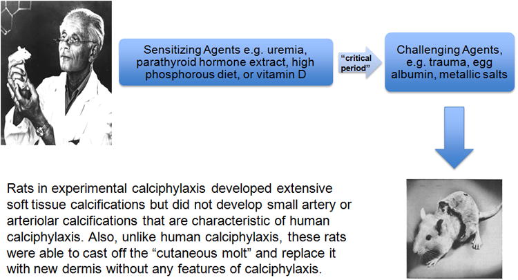
- Selye H, Gentile G, Prioreschi P. Cutaneous molt induced by calciphylaxis in the rat. Science. 1961;134(3493):1876–1877. - PubMed
-
- Skalnik J, Vasku J, Urbanek E. Calciphylaxis and cerebral atrophy in man. Clinical orthopaedics and related research. 1970;69:172–180. - PubMed
-
- Anderson DC, Stewart WK, Piercy DM. Calcifying panniculitis with fat and skin necrosis in a case of uraemia with autonomous hyperparathyroidism. Lancet. 1968;2(7563):323–325. - PubMed
-
- Budisavljevic MN, Cheek D, Ploth DW. Calciphylaxis in chronic renal failure. Journal of the American Society of Nephrology : JASN. 1996;7(7):978–982. - PubMed
Publication types
MeSH terms
Substances
Grants and funding
LinkOut - more resources
Full Text Sources
Other Literature Sources
Medical
