. 2015 May 11:6:7024.
doi: 10.1038/ncomms8024.

RNA structure determination by solid-state NMR spectroscopy

Affiliations

RNA structure determination by solid-state NMR spectroscopy

Alexander Marchanka et al. Nat Commun. .

Abstract

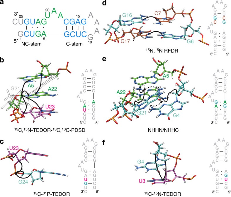
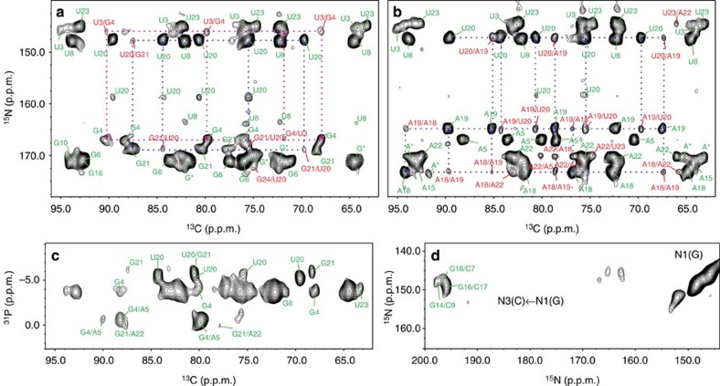
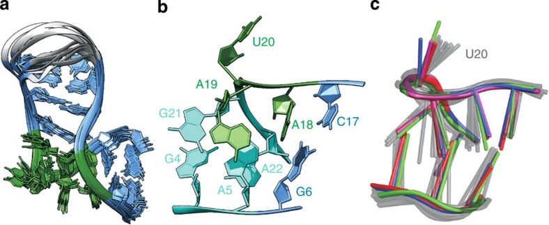
Knowledge of the RNA three-dimensional structure, either in isolation or as part of RNP complexes, is fundamental to understand the mechanism of numerous cellular processes. Because of its flexibility, RNA represents a challenge for crystallization, while the large size of cellular complexes brings solution-state NMR to its limits. Here, we demonstrate an alternative approach on the basis of solid-state NMR spectroscopy. We develop a suite of experiments and RNA labeling schemes and demonstrate for the first time that ssNMR can yield a RNA structure at high-resolution. This methodology allows structural analysis of segmentally labelled RNA stretches in high-molecular weight cellular machines—independent of their ability to crystallize—and opens the way to mechanistic studies of currently difficult-to-access RNA-protein assemblies.


Figures

Figure 1
Figure 1. Sequence of the Pf Box C/D RNA and magnetization transfer schemes.
(a) Sequence and secondary structure of the Pf Box C/D RNA. Helical regions, light blue; k-turn, green; loop and termini, grey. (bf) Schematic representation of the magnetization transfer schemes used for resonance assignment and distance measurement, shown on nucleotides stretches highlighted in the sequence. A, green; G, cyan; C, sienna; U, magenta. (b) 13C,15N-TEDOR-13C,13C-PDSD, A,Ulab-RNA. (c) 13C, 31P-TEDOR, G,Ulab-RNA. (d) 15N,15N-RFDR, G,Clab-RNA. (e) NHHN (dotted) and NHHC (solid), A,Glab-RNA. (f) 13C, 15N-TEDOR, (G-13C,U-15N)lab-RNA.
Figure 2
Figure 2. ssNMR spectra for the sequential assignment and measurement of structural restraints.
(ab) Ribose region of 2D 13C,15N-TEDOR-13C,13C-PDSD spectra of (a), G,Ulab-RNA and (b) A,Ulab-RNA (mixing time, 700 ms). Intra- and inter-nucleotide correlations are labeled in green and red, respectively. Selected sequential correlations are shown. Partially overlapped guanosines G10, G14, G16 are labeled as G*; non-site-specifically assigned adenosines in the tetra-loop (A11–A13) are labeled as A*. (c) 2D 13C,31P-TEDOR spectrum of G,Ulab-RNA. (d) 2D 15N,15N-RFDR spectrum showing the G-N1/C-N3 correlations for G:C base pairs.
Figure 3
Figure 3. ssNMR structure of the Pf Box C/D RNA.
(a) Overlay of the 10 lowest energy structures of the Pf Box C/D RNA in complex with L7Ae from ssNMR data. Terminal nucleotides 1 and 25–26 are not shown. Colour code as in Fig. 1a. (b) k-turn of the Pf Box C/D RNA, showing the characteristic geometry. Internal loop, green; NC stem, cyan, C stem, light blue. (c) Comparison of the k-turn geometry of the Pf Box C/D RNA obtained by ssNMR (10 lowest energy structures, gray) with that of the crystallographic structure of the Af Box C/D RNA (PDB code 1RLG), red; Pf Box C/D RNA (PDB code 3NMU), blue; Af Box C/D RNA (PDB code 4BW0), green; Ss Box C/D RNA (PDB code 3PLA), magenta.

References

    1. Petkova A. T. et al.. A structural model for Alzheimer's beta-amyloid fibrils based on experimental constraints from solid state NMR. Proc. Natl Acad. Sci. USA 99, 16742–16747 (2002). - PMC - PubMed
    1. Jaroniec C. P. et al.. High-resolution molecular structure of a peptide in an amyloid fibril determined by magic angle spinning NMR spectroscopy. Proc. Natl Acad. Sci. USA 101, 711–716 (2004). - PMC - PubMed
    1. de Groot H. J. M. Solid-state NMR spectroscopy applied to membrane proteins. Curr. Opin. Struct. Biol. 10, 593–600 (2000). - PubMed
    1. Opella S. J. & Marassi F. M. Structure determination of membrane proteins by NMR spectroscopy. Chem. Rev. 104, 3587–3606 (2004). - PMC - PubMed
    1. Shi L. C. et al.. Conformation of a seven-helical transmembrane photosensor in the lipid environment. Angew. Chem. Int. Ed. 50, 1302–1305 (2011). - PubMed

Publication types

Associated data