. 2015 Jun 2;23(6):1049-57.
doi: 10.1016/j.str.2015.03.020. Epub 2015 May 7.

Structural Basis for Multi-specificity of MRG Domains

Affiliations

Structural Basis for Multi-specificity of MRG Domains

Tao Xie et al. Structure. .

Abstract

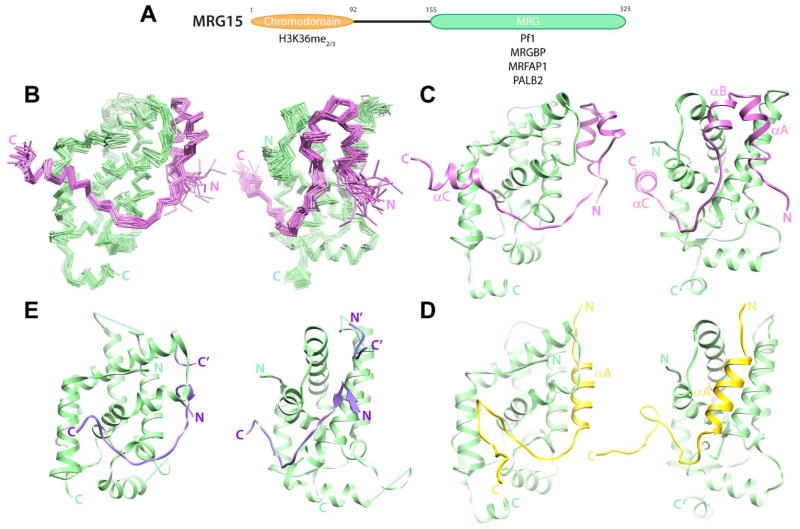
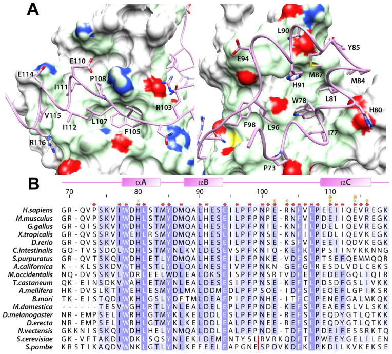
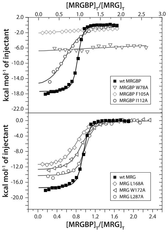
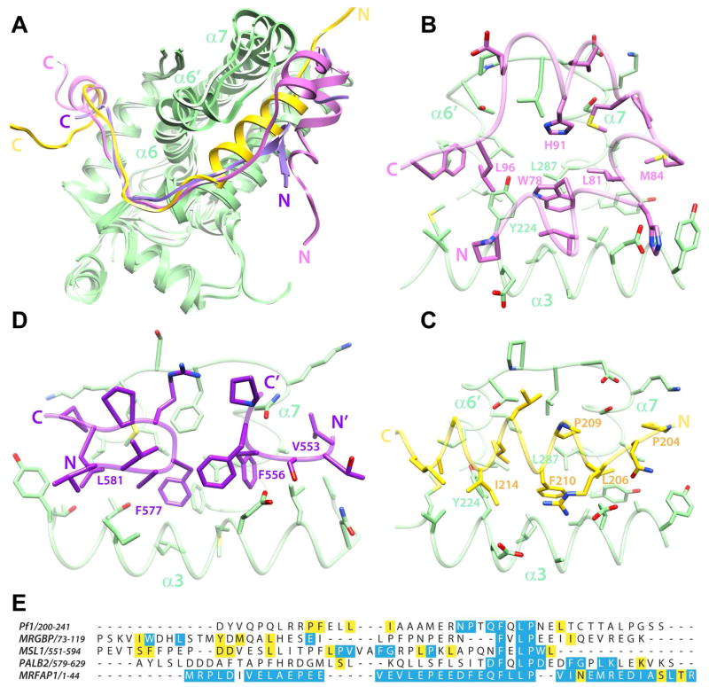
Chromatin-binding proteins play vital roles in the assembly and recruitment of multi-subunit complexes harboring effector proteins to specific genomic loci. MRG15, a chromodomain-containing chromatin-binding protein, recruits diverse chromatin-associated complexes that regulate gene transcription, DNA repair, and RNA splicing. Previous studies with Pf1, another chromatin-binding subunit of the Sin3S/Rpd3S histone deacetylase complex, defined the sequence and structural requirements for interactions with the MRG15 MRG domain, a common target of diverse subunits in the aforementioned complexes. We now show that MRGBP, a member of the Tip60/NuA4 histone acetyltransferase complex, engages the same two surfaces of the MRG domain as Pf1. High-affinity interactions occur via a bipartite structural motif including an FxLP sequence motif. MRGBP shares little sequence and structural similarity with Pf1, yet targets similar pockets on the surface of the MRG domain, mimicking Pf1 in its interactions. Our studies shed light onto how MRG domains have evolved to bind diverse targets.


Conflict of interest statement

The authors declare no conflicts of interest.

Figures

Figure 1
Figure 1
Structure of the human MRG15 MRG-MRGBP MBD complex and comparison with other protein-protein complexes involving MRG domains. (A) Domain structure of MRG15. Proteins and histone signals recognized by the individual domains are shown. (B) Backbone Cα traces of an ensemble of 20 NMR conformers following a best-fit superposition of secondary structural elements of MRG15 MRG (green) and MRGBP MBD (magenta). The two views are rotated by 70° to highlight the two MRG surfaces (Site I, right; Site II, left) engaged by MRGBP. (C) A representative conformer from the ensemble is shown with the backbone rendered as ribbons. Comparable views of (D) the MRG15 MRG-Pf1 MBD (PDB ID: 2LKM; (Xie et al., 2012)) and (E) the MSL3 MRG-MSL1 MBD (PDB ID: 2Y0N; (Kadlec et al., 2011)) complexes. Note that in the MSL3-MSL1 complex, two discrete MSL1 segments from molecules in adjacent complexes make contacts with Site I in the MRG domain; these segments are distinguished by N/C and N′/C′ labels. See also Figure S1 for biochemical data establishing the minimal MBD of MRGBP and Figure S2 for the quality of the NMR spectra obtained in the complex for both MRG15 MRG and MRGBP MBD.
Figure 2
Figure 2
Non-covalent interactions in the MRG15 MRG-MRGBP MBD complex. (A) Close-up views of the protein-protein interface highlighting the key residues within MRGBP MBD making contacts with the two MRG surfaces (Site I, right; Site II, left). The molecular surface of MRG domain in contact with MRGBP is rendered in green; the side chain nitrogen, sulfur, and oxygen atoms are colored blue, yellow, and red, respectively. (B) A multiple sequence alignment of MRGBP orthologs from a variety of species. The asterisks on top identify residues in the human protein involved in intermolecular interactions (red: hydrophobic; orange: hydrogen bonding; green: electrostatic). Residues are colored according to the degree of sequence conservation using JalView (Waterhouse et al., 2009). The red line denotes the location of multi-residue insertions in the yeast proteins. See also Figure S3 for the catalog of intermolecular interactions deduced using MONSTER (Salerno et al., 2004).
Figure 3
Figure 3
ITC analysis of binding of wild-type and various mutant MRG15 MRG and MRGBP MBD proteins. Representative binding curves from titrations of MRGBP MBD (in the syringe) with MRG15 MRG (in the cell) for the indicated proteins are shown. Titration data for mutant MRGBP MBD and MRG15 MRG are shown in the top and bottom panels, respectively. All experiments were conducted at 25 °C. See Supplementary Table S1 for the dissociation constants inferred from these measurements.
Figure 4
Figure 4
A comparative analysis of the structures of different MRG complexes. (A) Overlays of the MRG15 MRG-MRGBP MBD, MRG15 MRG-Pf1 MBD, and MSL3 MRG-MSL1 MBD complexes following best-fit superposition of backbone atoms in the MRG domains. The coloring scheme is the same as that in figure 1. Notice how the segment between α6′ and the N-terminus of α7 adopts different conformations in the three structures. This segment is relatively closed in the MSL1 complex but is progressively more open in the Pf1 and MRGBP complexes. Side chain interactions with various Site I residues in the respective MRG domains of the (B) MRGBP, (C) Pf1 and (D) MSL1 complexes are shown. The side chains of residues making comparable contacts in the various complexes are annotated. Note that in the MSL3-MSL1 complex, two discrete MSL1 segments from molecules in adjacent complexes make contacts with Site I in the MRG domain. (E) A CLUSTAL Ω-guided multiple sequence alignment of established MRG-interactors to emphasize the lack of sequence similarity between these domains. Conserved and invariant residues in the respective orthologous proteins are highlighted in yellow and blue, respectively. The high degree of sequence conservation for MRFAP1 is likely due to its occurrence in a narrow range of species. Notice the poor sequence conservation N-terminal to the FxLP motif in PALB2 orthologs. See also Figure S4 demonstrating a high-affinity interaction between PALB2 MBD and MRG15 MRG.

Similar articles

Cited by

References

    1. Bax A, Grzesiek S. Methodological advances in protein NMR. Accounts Chem Res. 1993;26:131–138.
    1. Bleuyard JY, Buisson R, Masson JY, Esashi F. ChAM, a novel motif that mediates PALB2 intrinsic chromatin binding and facilitates DNA repair. EMBO Rep. 2012;13:135–141. - PMC - PubMed
    1. Bowman BR, Moure CM, Kirtane BM, Welschhans RL, Tominaga K, Pereira-Smith OM, Quiocho FA. Multipurpose MRG domain involved in cell senescence and proliferation exhibits structural homology to a DNA-interacting domain. Structure. 2006;14:151–158. - PubMed
    1. Brünger AT, Adams PD, Clore GM, DeLano WL, Gros P, Grosse-Kunstleve RW, Jiang JS, Kuszewski J, Nilges M, Pannu NS, et al. Crystallography & NMR system: A new software suite for macromolecular structure determination. Acta Crystallogr D Biol Crystallogr. 1998;54:905–921. - PubMed
    1. Cai Y, Jin J, Tomomori-Sato C, Sato S, Sorokina I, Parmely TJ, Conaway RC, Conaway JW. Identification of new subunits of the multiprotein mammalian TRRAP/TIP60-containing histone acetyltransferase complex. J Biol Chem. 2003;278:42733–42736. - PubMed

Publication types

MeSH terms

Associated data

LinkOut - more resources