. 2015 Dec;11(12):1417-1429.
doi: 10.1016/j.jalz.2015.03.003. Epub 2015 May 7.

APOE effect on Alzheimer's disease biomarkers in older adults with significant memory concern

Affiliations

APOE effect on Alzheimer's disease biomarkers in older adults with significant memory concern

Shannon L Risacher et al. Alzheimers Dement. 2015 Dec.

Abstract

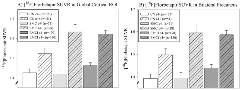
Introduction: This study assessed apolipoprotein E (APOE) ε4 carrier status effects on Alzheimer's disease imaging and cerebrospinal fluid (CSF) biomarkers in cognitively normal older adults with significant memory concerns (SMC).

Methods: Cognitively normal, SMC, and early mild cognitive impairment participants from Alzheimer's Disease Neuroimaging Initiative were divided by APOE ε4 carrier status. Diagnostic and APOE effects were evaluated with emphasis on SMC. Additional analyses in SMC evaluated the effect of the interaction between APOE and [(18)F]Florbetapir amyloid positivity on CSF biomarkers.

Results: SMC ε4+ showed greater amyloid deposition than SMC ε4-, but no hypometabolism or medial temporal lobe (MTL) atrophy. SMC ε4+ showed lower amyloid beta 1-42 and higher tau/p-tau than ε4-, which was most abnormal in APOE ε4+ and cerebral amyloid positive SMC.

Discussion: SMC APOE ε4+ show abnormal changes in amyloid and tau biomarkers, but no hypometabolism or MTL neurodegeneration, reflecting the at-risk nature of the SMC group and the importance of APOE in mediating this risk.

Keywords: ADNI; APOE; Alzheimer's Disease Neuroimaging Initiative; Apolipoprotein E; CSF; Cerebrospinal fluid; FDG; Neuroimaging; PET; SCD; SMC; Significant memory concern; Structural magnetic resonance imaging (MRI); Subjective cognitive decline; [(18)F]Florbetapir PET; [(18)F]Fluorodeoxyglucose.


Figures

Figure 1
Figure 1. Impact of diagnosis and APOE ε4 carrier status on cerebral amyloid deposition
(A) Voxel-wise analysis of [18F]Florbetapir PET scans showed a main effect of APOE ε4 carrier status such that APOE ε4+ participants had greater amyloid deposition than APOE ε4− participants in nearly the entire cortex. (B) A main effect of diagnostic group (EMCI>SMC>CN) was also observed in more restricted regions of the frontal, temporal, and medial parietal cortices. Significant effects of APOE ε4 carrier status within diagnostic groups were also observed, including in (C) CN participants (127 CN ε4−, 51 CN ε4+), (D) SMC participants (71 SMC ε4−, 28 SMC ε4+), and (E) EMCI participants (170 EMCI ε4−, 130 EMCI ε4+) in widespread cortical regions, including in the frontal, parietal, temporal, and occipital lobes. No regions showed higher amyloid deposition in APOE ε4 non-carriers than APOE ε4 carriers. Figure is displayed at voxel-wise threshold of p<0.001 (uncorrected for multiple comparisons); minimum voxel size (k) = 300 voxels, which corresponds to a cluster-wise threshold of p<0.05 (family-wise error (FWE) correction for multiple comparison).
Figure 2
Figure 2. Impact of diagnosis and APOE ε4 carrier status on regional amyloid deposition
Regional analysis of amyloid deposition on [18F]Florbetapir PET showed that APOE ε4+ participants had greater amyloid deposition than APOE ε4− participants in (A) a global cortical region of interest, as well as within the (B) bilateral precuneus, across all diagnostic groups (APOE ε4 carrier status: both p<0.001). Diagnostic group was also significantly associated with amyloid deposition in both target regions (diagnosis: both p<0.01). In Bonferroni-corrected post-hoc pair comparisons, SMC and EMCI ε4+ participants showed higher amyloid than CN ε4−, SMC ε4−, and EMCI ε4− participants in both ROIs (all p<0.05), CN ε4+ participants had higher amyloid than CN ε4− in the global cortex (A; p=0.052) and bilateral precuneus (B; p<0.05), and EMCI ε4+ participants had greater amyloid deposition in the global cortical ROI than CN ε4+ participants (A; p<0.05). No significant interaction effect of diagnostic group and APOE ε4 carrier status was observed, although a trend for an interaction effect on global cortical amyloid deposition was observed (A; p=0.084).
Figure 3
Figure 3. Relationship of diagnosis and APOE ε4 carrier status with regional glucose metabolism and medial temporal atrophy
The effect of diagnostic group and APOE ε4 carrier status on (A) global cortical or (B) parietal lobe glucose metabolism, (C) hippocampal volume, and (D) entorhinal cortex thickness was evaluated. No significant independent effect of APOE ε4 carrier status on across CN, SMC, or EMCI participants in any measure interest was observed, although diagnosis was significant for global hypometabolism region (A; p=0.001), mean parietal hypometabolism (B; p=0.048), and hippocampal (C; p<0.001) and entorhinal cortex (D; p=0.003) atrophy. A significant interaction effect on hypometabolism, but not atrophy, between diagnosis and APOE ε4 carrier status was observed (global cortex: p=0.014; mean parietal lobe: p=0.016). Post-hoc analyses showed that EMCI ε4+ participants had reduced glucose metabolism relative CN ε4−, SMC ε4−, SMC ε4+, and EMCI ε4− in the global cortical region (A; all p<0.05) and relative to EMCI ε4− participants only in the mean parietal lobe (B; p<0.05). Further, post-hoc comparisons showed that (C) hippocampal volume was significantly reduced in EMCI participants regardless of APOE ε4 carrier status relative to CN ε4− and SMCI ε4+ participants, as well as in EMCI ε4− participants relative to CN ε4+ participants (all p<0.05). (D) Entorhinal cortex thickness was reduced in EMCI ε4− relative to CN ε4− participants (p<0.001).
Figure 4
Figure 4. Relationship of diagnosis and APOE ε4 carrier status with CSF protein levels
Diagnostic group and APOE ε4 carrier status were significantly associated with CSF levels of (A) Aβ1–42, (B) t-tau, and (C) phosphorylated tau (p-tau). (A) CSF Aβ1–42 levels showed a significant independent effect of both diagnostic group (p=0.023) and APOE ε4 carrier status (p<0.001) but no interaction effect. On post-hoc analysis, ε4+ participants showed lower CSF Aβ1–42 than ε4− participants regardless of diagnostic group (i.e., CN ε4−, SMC ε4−, EMCI ε4− > CN ε4+, SMC ε4+, EMCI ε4+; all p<0.05). (B) Significant independent effects of diagnosis and APOE ε4 carrier status (p<0.001), as well as an interaction effect between diagnostic group and APOE ε4 carrier status (p=0.002), on CSF t-tau were also observed. EMCI ε4+ participants showed higher t-tau levels than CN ε4−, CN ε4+, SMC ε4−, and EMCI ε4− participants (all p<0.05) on post-hoc analysis. (C) CSF p-tau level was significantly associated with APOE ε4 carrier status only (p<0.001). On post-hoc analysis, EMCI and SMC ε4+ participants showed higher p-tau levels than CN, SMC, and EMCI ε4− participants (all p<0.05).
Figure 5
Figure 5. Effect of APOE ε4 carrier status and amyloid positivity on CSF biomarkers in SMC participants
(A) Significant independent effects of APOE ε4 carrier status and cerebral amyloid status (positive or negative), but no interaction effect, on CSF Aβ1–42 were observed (both p<0.001). Post-hoc analysis indicated a pattern of decreasing CSF level Aβ1–42 by the interaction of amyloid positivity and APOE ε4+ was seen with SMC APOE ε4−/Aβ− showing higher levels than SMC APOE ε4+/Aβ+, SMC APOE ε4+/Aβ−, and SMC APOE ε4−/Aβ+, and SMC APOE ε4+/Aβ− showing higher CSF Aβ1–42 levels than SMC APOE e4+/Aβ+ (all p<0.05). (B) APOE ε4 carrier status, but not amyloid positivity or the interaction, was significantly associated with CSF t-tau level in the SMC participants (p=0.009). SMC APOE ε4+/Aβ+ showed higher t-tau levels than SMC APOE ε4−/Aβ− on Bonferroni-corrected post-hoc analysis ( p=0.008). (C) Both APOE ε4 carrier status and amyloid positivity were independently associated with CSF p-tau level (APOE ε4 carrier status, p = 0.014; amyloid positivity, p = 0.010), but again no interaction was observed. Similar to the t-tau analyses, SMC APOE ε4+/Aβ+ had greater CSF p-tau levels than SMC APOE ε4−/Aβ− on post-hoc analysis (p<0.001).

Similar articles

Cited by

References

    1. Alzheimer’s Association. 2014 Alzheimer’s disease facts and figures. Alzheimers Dement. 2014;10:e47–e92. - PubMed
    1. Sperling RA, Aisen PS, Beckett LA, Bennett DA, Craft S, Fagan AM, et al. Toward defining the preclinical stages of Alzheimer’s disease: recommendations from the National Institute on Aging-Alzheimer’s Association workgroups on diagnostic guidelines for Alzheimer’s disease. Alzheimers Dement. 2011;7:280–92. - PMC - PubMed
    1. Jessen F, Amariglio RE, van Boxtel M, Breteler M, Ceccaldi M, Chetelat G, et al. A conceptual framework for research on subjective cognitive decline in preclinical Alzheimer’s disease. Alzheimers Dement. 2014 - PMC - PubMed
    1. Saykin AJ, Wishart HA, Rabin LA, Santulli RB, Flashman LA, West JD, et al. Older adults with cognitive complaints show brain atrophy similar to that of amnestic MCI. Neurology. 2006;67:834–42. - PMC - PubMed
    1. Caselli RJ, Chen K, Locke DE, Lee W, Roontiva A, Bandy D, et al. Subjective cognitive decline: self and informant comparisons. Alzheimers Dement. 2014;10:93–8. - PMC - PubMed

Publication types

MeSH terms