

Periapical lesions of the jaws: a review of 104 cases in ibadan
- PMID: 25960702
- PMCID: PMC4415388
Periapical lesions of the jaws: a review of 104 cases in ibadan
Abstract
Background: Periapical lesions (PLs) occur as a result of pulpal inflammation and may rarely be seen in the absence of pulpal diseases. They are the most common pathological lesions affecting the alveolar bone.
Objective: This study aims to describe the clinicopathological features of PLs of the jaws with emphasis on the two most common types.
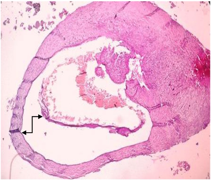
Methods: Histopathology records of PLs diagnosed from January 1990 to December 2012 at the Department of Oral Pathology, University College Hospital Ibadan, were examined and categorized into periapical cysts (PCs); periapical granuloma (PGs) and others. Clinical data and histopathological features of these PLs were reviewed and analyzed.
Results: One hundred and four lesions met the criteria for this study and consisted of PGs with 71 (68.3%) cases and PCs with 31 (29.8%) cases and one case each of apical scar and pleomorphic adenoma. Age range of cases was 9 to 80 years (mean=35.6 ± 15.8years) with a peak at age group of 20-29 years. Females were more frequently affected with 51.9% of cases. PLs were most frequently diagnosed in the anterior maxillary region with 58 (56.9%) cases, while the most frequently involved tooth was the left maxillary central incisor with 23 (22.1%) cases.
Conclusion: Findings in this study are consistent with those of previous studies. It is important for all periapical pathological specimens to be submitted for histological examination to establish an accurate diagnosis and aid in the identification of sinister lesions that may present in the Periradicular region of teeth.
Keywords: Cyst; Granuloma; Histopathology; Jaw; Periapical.
Figures


References
-
- Cotti E, Campisi G. Advanced radiographic techniques for the detection of lesions in bone. Endod Topics. 2004;7: 52–72.
-
- Abbott PV. Classification, diagnosis and clinical manifestations of apical periodontitis. Endod Topics. 2004;8: 36–54.
-
- Peters E, Lau M. Histopathologic examination to confirm diagnosis of periapical lesions: a review. J Can Dent Assoc. 2003;69(9):598–600. - PubMed
-
- Ramanpreet B, Simarpreet SV, Rajat B , et al. Histopathological insight into periapical lesions: an institutional study from Punjab. Int J Oral Maxillofac Pathol. 2012;3(3):02–07.
Publication types
LinkOut - more resources
Full Text Sources
Miscellaneous