

Detection of tropical fungi in formalin-fixed, paraffin-embedded tissue: still an indication for microscopy in times of sequence-based diagnosis?
- PMID: 25961048
- PMCID: PMC4417575
- DOI: 10.1155/2015/938721
Detection of tropical fungi in formalin-fixed, paraffin-embedded tissue: still an indication for microscopy in times of sequence-based diagnosis?
Abstract
Introduction: The aim of the study was the evaluation of panfungal PCR protocols with subsequent sequence analysis for the diagnostic identification of invasive mycoses in formalin-fixed, paraffin-embedded tissue samples with rare tropical mycoses.
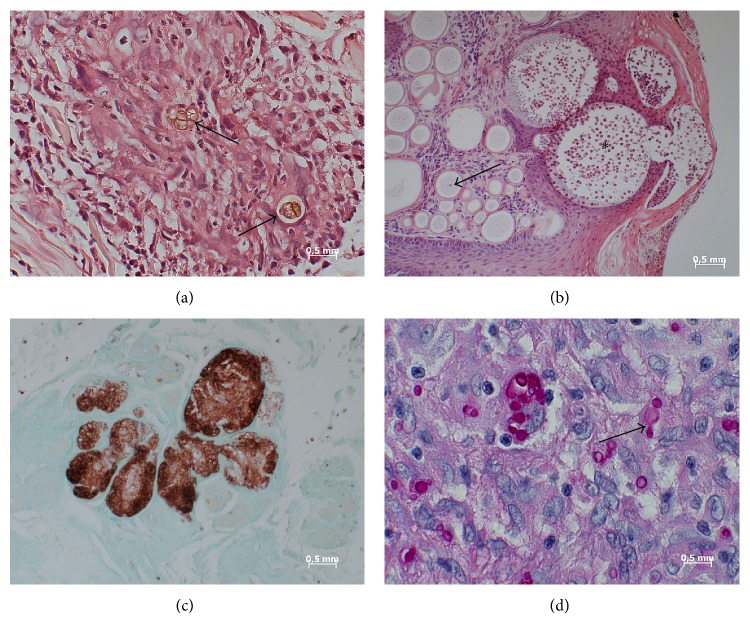
Materials and methods: Five different previously described panfungal PCR/sequencing protocols targeting 18S and 28S ribosomal RNA gene fragments as well as internal transcribed spacer 1 and 2 fragments were evaluated with a collection of 17 formalin-fixed, paraffin-embedded tissue samples of patients with rare and/or tropical invasive mycoses, comprising chromoblastomycosis, coccidioidomycosis, cryptococcosis, histoplasmosis, mucormycosis, mycetoma/maduromycosis, and rhinosporidiosis, in a proof-of-principle analysis.
Results: The primers of the panfungal PCRs readily and predominantly reacted with contaminating environmental fungi that had deposited on the paraffin blocks. Altogether three sequence results of histoplasmosis and mycetoma samples that matched the histological assessment were associated with sample age <10 years and virtually without PCR inhibition.
Conclusions: The high risk of amplifying environmental contaminants severely reduces the usefulness of the assessed panfungal PCR/sequencing protocols for the identification of rare and/or tropical mycoses in stored formalin-fixed, paraffin-embedded tissues. Histological assessment remains valuable for such indications if cultural differentiation is impossible from inactivated sample material.
Figures
References
-
- Sangoi A. R., Rogers W. M., Longacre T. A., Montoya J. G., Baron E. J., Banaei N. Challenges and pitfalls of morphological identification of fungal infections in histologic and cytologic specimens. A ten-year retrospective review at a single institution. The American Journal of Clinical Pathology. 2009;131(3):364–375. doi: 10.1309/ajcp99ooozsniscz. - DOI - PubMed
-
- Watts J. C. Surgical pathology and the diagnosis of infectious diseases. American Journal of Clinical Pathology. 1994;102(6):711–712. - PubMed
Publication types
MeSH terms
Substances
LinkOut - more resources
Full Text Sources
Other Literature Sources
Medical
Molecular Biology Databases
