. 2015 Jun;17(6):782-92.
doi: 10.1038/ncb3170. Epub 2015 May 11.

A nuclear role for the respiratory enzyme CLK-1 in regulating mitochondrial stress responses and longevity

A nuclear role for the respiratory enzyme CLK-1 in regulating mitochondrial stress responses and longevity

Richard M Monaghan et al. Nat Cell Biol. 2015 Jun.

Abstract

The coordinated regulation of mitochondrial and nuclear activities is essential for cellular respiration and its disruption leads to mitochondrial dysfunction, a hallmark of ageing. Mitochondria communicate with nuclei through retrograde signalling pathways that modulate nuclear gene expression to maintain mitochondrial homeostasis. The monooxygenase CLK-1 (human homologue COQ7) was previously reported to be mitochondrial, with a role in respiration and longevity. We have uncovered a distinct nuclear form of CLK-1 that independently regulates lifespan. Nuclear CLK-1 mediates a retrograde signalling pathway that is conserved from Caenorhabditis elegans to humans and is responsive to mitochondrial reactive oxygen species, thus acting as a barometer of oxidative metabolism. We show that, through modulation of gene expression, the pathway regulates both mitochondrial reactive oxygen species metabolism and the mitochondrial unfolded protein response. Our results demonstrate that a respiratory enzyme acts in the nucleus to control mitochondrial stress responses and longevity.


Figures

Figure 1
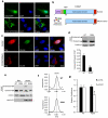
Figure 1. CLK-1 and its human homologue COQ7 localise to mitochondria and nuclei
(a) Endogenous COQ7 and COXIV (mitochondrial marker) immunostaining in HeLa cells. Nuclei stained with DAPI. (b) HeLa cells expressing COQ7 tagged at the C-terminus with Myc epitope (COQ7-Myc) were stained with anti-Myc and anti-COXIV antibodies. Quantification of nuclear COQ7-Myc staining is in Supplementary Fig. 1a. (c) Wild type CLK-1 (CLK-1wt) fused at the C-terminus to GFP localises to both mitochondria and nuclei in adult C. elegans. Arrow marks nucleus. MT (MitoTracker, mitochondrial marker). (d) HeLa cells immunostained with an antibody specific to the N-terminus (amino acids 1-37) of COQ7 (COQ7N-term). (e) siRNA targeting COQ7 transcripts decrease levels of both cleaved and uncleaved COQ7 protein. Immunoblots of lysates from HEK293 cells transfected with non-targeting (CTRL) or COQ7 siRNA. Short and long exposures are shown. (f) Quantification of nuclear staining intensity of cells immunostained with anti-COQ7(1-37) (COQ7N-term) following treatment with antioxidant (N-acetyl cysteine, NAC, 10 mM, 24 h) or exogenous ROS (hydrogen peroxide, 200 μM or 800 μM, respectively, 3 h) compared to untreated (0) control. 50 cells assessed per experiment in n=3 independent experiments (error bars, s.e.m. *P < 0.05). (g) Percent of mCherry-positive nuclei that are also GFP-positive in C. elegans expressing CLK-1-GFP and the nuclear marker HIS-24-mCherry. 25 worms were assessed per experiment in n=3 independent experiments (error bars, s.e.m. **P < 0.005). (h) The ratio of uncleaved to cleaved COQ7 in lysates from HEK293 was quantified from n=3 independent immunoblotting experiments (error bars, s.e.m. *P < 0.05, **P < 0.005). Cells were treated with hydrogen peroxide (H2O2, 150 μM, 3h; cellular ROS) or rotenone (50 μM, 3 h; complex I inhibitor, mitochondrial ROS), with or without 10 mM NAC. A representative immunoblot is shown. (i) Immunoblots of lysates from HEK293 cells expressing COQ7-Myc or COQ7 (R11/14/16D)-Myc (mutant that disrupts mitochondrial targeting) treated with H2O2 (150 μM, 4 h) or NAC (10 mM, 6 h). NT = untreated cells. (j) Blocking mitochondrial targeting of COQ7 enhances nuclear localisation of uncleaved COQ7. HeLa cells expressing a C-terminally Myc-tagged COQ7 (R11/14/16D) mutant, that disrupts mitochondrial targeting, were immunostained with anti-Myc and anti-COXIV antibodies. Uncropped images of immunoblots are shown in Supplementary Fig. 5.
Figure 2
Figure 2. Nuclear COQ7 functions independently of the mitochondrial form
(a) Reduced nuclear localisation of COQ7 (R28A) mutant. Fluorescence in COS7 cells expressing COQ7 or COQ7 (R28A) fused at the C-terminus to GFP. Nuclei stained with DAPI. (b) Schematic depicting location of the mitochondrial targeting sequence (MTS), the nuclear targeting sequence (NTS) and the mitochondrial processing peptidase cleavage site (MPP) on COQ7. The N-terminal region of COQ7 is degraded following cleavage by MPP in mitochondria. Also shown are the positions of the OLLAS and FLAG epitope-tags. Numbers refer to amino acid positions of COQ7. (c) HeLa cells expressing dual OLLAS and FLAG tagged COQ7 or COQ7 (R28A) immunostained with anti-FLAG and anti-OLLAS antibodies. The anti-FLAG antibodies recognise total COQ7 (uncleaved and cleaved) and the anti-OLLAS antibody specifically recognises uncleaved nuclear COQ7. (d) Immunoblot of cell lysates from HEK293 cells stably expressing either untagged wild type (WT) COQ7 or the R28A mutant in the presence of siRNA against untranslated regions of COQ7. Quantification of the ratio of uncleaved (nuclear) to cleaved (mitochondrial) COQ7 from n=3 independent experiments is presented (error bars, s.e.m. **P < 0.005). (e) Immunoblot of cell lysates from the stable HEK293 cells expressing either wild type (WT) COQ7 or the R28A mutant separated into mitochondrial (Mito) and nuclear (Nuclei) pellet fractions (COXIV, mitochondrial marker; Lamin B1, nuclear matrix marker). The presence of some COXIV and cleaved COQ7 in the nuclear fraction indicates some mitochondrial contamination is present. (f) Ubiquinone (UQ10) levels are similar in COQ7 WT and R28A expressing cells. Reverse phase HPLC chromatograms of quinones purified from cells (UQ10 peak at 8.63 minutes). (g) Proliferation of COQ7 (R28A) expressing cells was reduced compared to COQ7 WT cells in the presence of siRNA targeting endogenous COQ7 (siCOQ7) but not control siRNA (siCTRL). Measured by MTT assay (mean values from 4 wells of cells per condition in n=3 independent experiments; error bars, s.e.m. *P < 0.05). Cell survival was not altered under these conditions (see Supplementary Fig. 2f). Uncropped images of immunoblots are shown in Supplementary Fig. 5.
Figure 3
Figure 3. A truncated form of C. elegans CLK-1 with impaired mitochondrial targeting is predominantly nuclear and does not rescue ubiquinone biosynthesis
(a) The mechanism of targeting of C. elegans CLK-1 and human COQ7 to nuclei is not conserved. Alignment of the N-termini and the start of the highly conserved diiron binding domains of CLK-1 and COQ7. The mitochondrial targeting sequence (MTS) is denoted by the black bars, conserved amino acids are highlighted in blue, and the predicted mitochondrial processing peptidase (MPP) site in COQ7 is shown. The region in COQ7 containing determinants of nuclear localisation (NTS) is denoted by the red bar and residues required for nuclear-targeting are highlighted in red. CE, C. elegans; HS, Homo sapiens. Numbers refer to amino acid positions. (b) CLK-1 lacking the MTS (CLK-1nuc(+)) is predominantly nuclear in adult worms. Arrows mark nuclei. MT (MitoTracker, mitochondrial marker). (c) CLK-1nuc(+) expression does not rescue the loss of mitochondrial ubiquinone (UQ9) biosynthesis in clk-1(−) worms. Reverse phase HPLC chromatograms of quinones extracted from the indicated strains (UQ9 peak at 8.78 minutes, DMQ9 peak at 8.42 minutes). CLK-1wt but not CLK-1nuc(+) restores the UQ9 peak lost in clk-1 (clk-1(−)) null worms.
Figure 4
Figure 4. Nuclear CLK-1/COQ7 regulates ROS metabolism
(a) Nuclear CLK-1 acts to lower cellular ROS levels. Expression of the nuclear form of CLK-1 (clk-1nuc(+)) in clk-1 null worms (clk-1(−)) partially rescues the increased levels of ROS observed in these worms measured using the ROS-sensitive dye DHE. Wild-type CLK-1-GFP (clk-1wt) completely rescues the phenotype. Representative images and quantification of ROS levels are shown (25 worms assessed per experiment in n=3 independent experiments; error bars, s.e.m. **P < 0.005, ***P < 0.001). (b) Expression of CLK-1nuc(+) in clk-1 null worms significantly increases their survival in response to treatment with the respiratory inhibitor paraquat (40 mM, 6 h). CLK-1wt expression in clk-1 null worms rescues survival to levels similar to N2 worms (25 worms assessed per experiment in n=3 independent experiments; error bars, s.e.m. **P < 0.01). (c) Increased levels of ROS in untreated and oxidative stress treated (100 μM tert-butyl hydroperoxide, 1 h) HEK293 cells treated with COQ7 siRNA and expressing non-nuclear COQ7 (R28A) compared to wild type (WT), monitored by DCF fluorescence (mean values from 4 wells of cells per condition in n=3 independent experiments; error bars, s.e.m. *P < 0.05, **P < 0.005). (d) Nuclear COQ7 promotes resistance to ROS insult. Increased sensitivity of R28A expressing cells to oxidative stress (1 mM CoCl2, 4 h) measured by MTT assay (mean values from 4 wells of cells per condition in n=3 independent experiments; error bars, s.e.m. *P < 0.05). (e, f) Nuclear CLK-1 regulates the expression of genes involved in ROS metabolism. Altered transcript levels of glna-1 and dhs-7 in clk-1 null worms (clk-1(−)) are rescued by expression of CLK-1nuc(+) (mean values from 3 reactions per condition for n=3 independent experiments; error bars, s.e.m. *P < 0.05). (g) The transcript levels of the GLS2 and WWOX are decreased or increased, respectively, upon loss of nuclear COQ7 (R28A compared to WT) (mean values from 3 reactions per condition for n=4 independent experiments; error bars, s.e.m. *P < 0.05, **P < 0.005). (h) The transcript levels of HTRA2 are increased in cells lacking nuclear COQ7 (R28A compared to WT) (mean values from 3 reactions per condition for n=4 independent experiments; error bars, s.e.m. **P < 0.005). (i) Immunoblots of corresponding protein levels for gene transcripts analysed in Fig. 4g, h and Supplementary Fig. 3c (NRF2). Uncropped images of blots are shown in Supplementary Fig. 5. (j) Inhibition of HTRA2 activity (10 mM UCF-101, 30 min) rescues the ROS sensitivity of COQ7 (R28A) cells following oxidative stress (1 mM CoCl2, 4 h). Cell survival measured by MTT assay relative to DMSO treated cells (mean values from 4 wells of cells per condition in n=3 independent experiments; error bars, s.e.m. *P < 0.05).
Figure 5
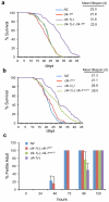
Figure 5. Mitochondrial and nuclear CLK-1 independently contribute to longevity
(a, b) CLK-1nuc(+) expression partially rescues the increased lifespan observed in clk-1(−) worms while CLK-1wt completely rescues the longevity phenotype. Lifespan plotted as percent survival and mean lifespans calculated. N2 is the wild type strain. Lifespan data, including mean, maximum and 90th percentile lifespan with statistical analysis, for n=3 independent experiments is reported in Supplementary Table 1. (c) Analysis of developmental timing. Worms were synchronised at L1 and larval stage was determined every 24 hours until all of the worms reached adulthood. Approximately 50 worms per genotype were monitored for each time point (mean values from n=3 independent experiments; error bars, s.e.m.).
Figure 6
Figure 6. Nuclear CLK-1/COQ7 suppresses the expression of a subset of UPRmt genes
(a) CLK-1nuc(+) expressing worms display decreased hsp-6::gfp reporter activity compared to clk-1(−) worms. The unmodified hsp-6::gfp reporter strain is designated clk-1(+). Quantification of reporter fluorescence in CLK-1nuc(+) expressing worms (clk-1(−); clk-1nuc(+)) relative to clk-1(−) worms (mean fluorescence of 50 worms per genotype pooled from n=3 independent experiments; error bars, s.e.m. **P < 0.005). (b) qPCR measuring mRNA transcripts of UPRmt genes in clk-1(−) or clk-1(−); clk-1nuc(+) worms relative to wild type strain (N2) (mean values from 3 reactions per condition in n=3 independent experiments; error bars, s.e.m., n.s., no significant difference, *P < 0.05). (c) Heat map depicting change in expression of UPRmt genes (MT) and UPRER (ER) genes in R28A cells compared to WT COQ7 cells. Map generated from the qPCR data presented in Supplementary Figure 4b and is representative of n=3 independent experiments. Scale represents mean fold change in expression. (d) Immunoblots of levels of UPRmt proteins including the mitochondrial controls COXIV (nuclear-encoded) and MTCO1 (mitochondrial-encoded). Uncropped images of blots are shown in Supplementary Fig. 5.
Figure 7
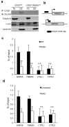
Figure 7. COQ7 associates with chromatin
(a) Chromatin fractionation of HEK293 cells expressing wild type or R28A (non-nuclear mutant) COQ7 tagged with an internal OLLAS epitope (as shown in Fig. 2b) followed by anti-COQ7 immunoprecipitation and anti-OLLAS immunoblot. Each fraction was also immunoblotted for the markers Tubulin β (cytosolic), c-Jun (active chromatin), Lamin B1 (nuclear matrix). TS, triton-soluble fraction; DS, DNase-soluble fraction. (b) Schematic of the WWOX and TIMM22 COQ7-associated promoter sites enriched in anti-COQ7 ChIPs compared to IgG control. Arrows denote transcriptional start sites. Full data set of enriched sites is provided in Supplementary Table 2. (c) Anti-COQ7 ChIP was performed on HEK293 cells expressing wild type (WT) or the non-nuclear form (R28A) of COQ7 before qPCR analysis of WWOX and TIMM22 promoter sites and two control intergenic sites (CTRL1 and CTRL2) (mean values from 3 reactions per condition in n=3 independent experiments; error bars, s.e.m. n.s., no significant difference; **P < 0.005). (d) Anti-COQ7 ChIP was performed on HEK293 cells treated with antioxidant (N-acetyl cysteine, NAC, 10 mM, 24 h) compared to untreated (mean values from 3 reactions per condition in n=3 independent experiments; error bars, s.e.m. n.s., no significant difference; *P < 0.05).
Figure 8
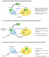
Figure 8. Model for the regulation of ROS metabolism, the UPRmt and lifespan by nuclear CLK-1/COQ7
(a) CLK-1/COQ7 regulates mitochondrial homeostasis. The majority of CLK-1/COQ7 localises to mitochondria by means of its mitochondrial targeting sequence (MTS), where it is required for the biosynthesis of ubiquinone (UQ), an essential cofactor in the electron transport chain (ETC). However, basal levels of ROS, produced by the mitochondria, direct a pool of CLK-1/COQ7 to the nucleus where it regulates gene expression. Some CLK-1/COQ7-regulated genes are directly involved in mitochondrial ROS metabolism and, therefore, the prolonged presence of CLK-1/COQ7 in the nucleus lowers ROS levels. Reduced ROS leads to CLK-1/COQ7 being predominantly localised to mitochondria, and not the nucleus, so its affects on gene expression are relieved, basal ROS production returns, and homeostasis is maintained. (b) Loss of nuclear COQ7 (R28A mutant) in human cells or loss of CLK-1 in worms (that scavenge UQ from their bacterial diet) alters ROS metabolism leading to increased ROS levels, augments the UPRmt, and extends the lifespan of worms. (c) The augmented ROS levels, UPRmt and extended lifespan in clk-1(−) worms is suppressed by expression of a nuclear-localised CLK-1 mutant (CLK-1nuc(+)) that acts to try and maintain mitochondrial homeostasis.

Similar articles

Cited by

References

    1. Tait SW, Green DR. Mitochondria and cell signalling. J. Cell Sci. 2012;125:807–815. - PMC - PubMed
    1. Chandel NS. Mitochondria as signaling organelles. BMC Biol. 2014;12:34. - PMC - PubMed
    1. Sena LA, Chandel NS. Physiological roles of mitochondrial reactive oxygen species. Mol. Cell. 2012;48:158–167. - PMC - PubMed
    1. Whelan SP, Zuckerbraun BS. Mitochondrial signaling: forwards, backwards, and in between. Oxid. Med. Cell. Longev. 2013;2013:351613. - PMC - PubMed
    1. Kotiadis VN, Duchen MR, Osellame LD. Mitochondrial quality control and communications with the nucleus are important in maintaining mitochondrial function and cell health. Biochim. Biophys. Acta. 2014;1840:1254–1265. - PMC - PubMed

Publication types

MeSH terms