. 2015 May 11;10(5):e0125962.
doi: 10.1371/journal.pone.0125962. eCollection 2015.

Chaperone-Mediated Autophagy Targets IFNAR1 for Lysosomal Degradation in Free Fatty Acid Treated HCV Cell Culture

Affiliations

Chaperone-Mediated Autophagy Targets IFNAR1 for Lysosomal Degradation in Free Fatty Acid Treated HCV Cell Culture

Ramazan Kurt et al. PLoS One. .

Expression of concern in

Abstract

Background: Hepatic steatosis is a risk factor for both liver disease progression and an impaired response to interferon alpha (IFN-α)-based combination therapy in chronic hepatitis C virus (HCV) infection. Previously, we reported that free fatty acid (FFA)-treated HCV cell culture induces hepatocellular steatosis and impairs the expression of interferon alpha receptor-1 (IFNAR1), which is why the antiviral activity of IFN-α against HCV is impaired.

Aim: To investigate the molecular mechanism by which IFNAR1 expression is impaired in HCV cell culture with or without free fatty acid-treatment.

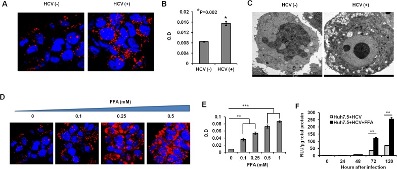
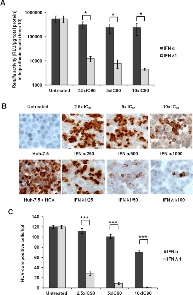
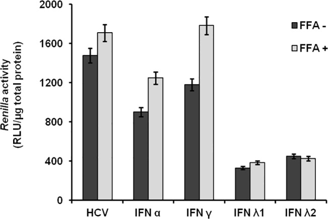
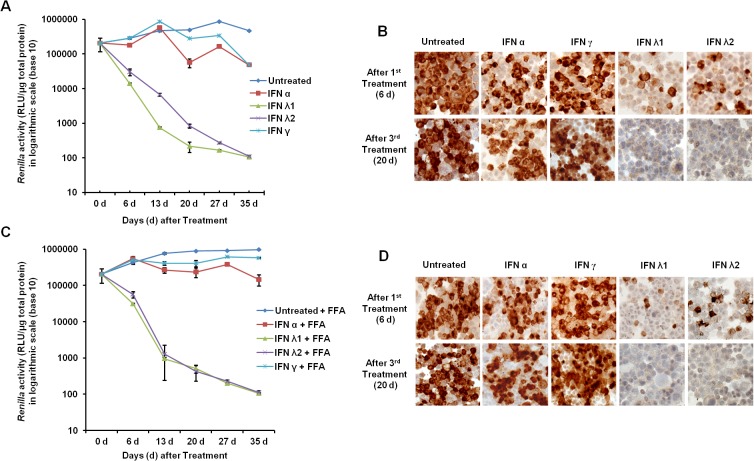
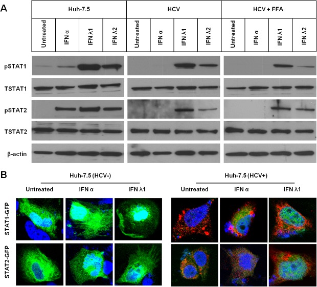
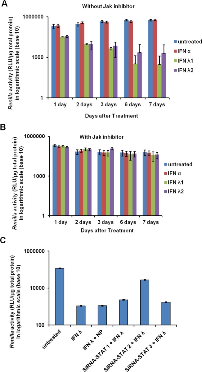
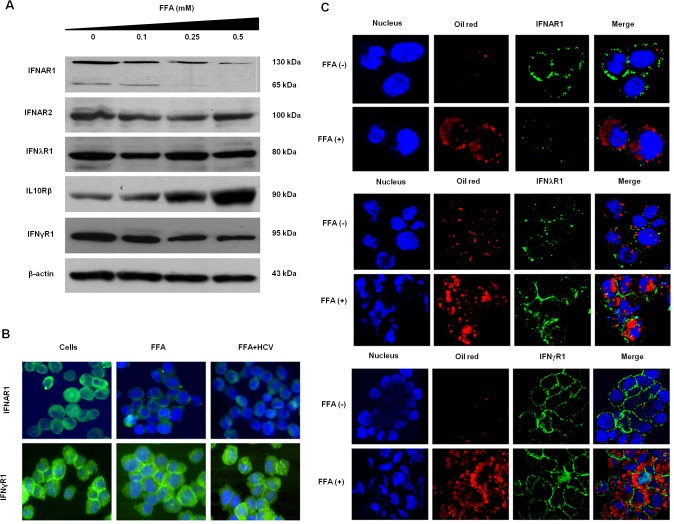
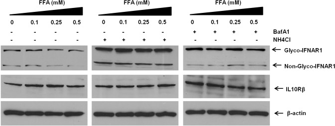
Method: HCV-infected Huh 7.5 cells were cultured with or without a mixture of saturated (palmitate) and unsaturated (oleate) long-chain free fatty acids (FFA). Intracytoplasmic fat accumulation in HCV-infected culture was visualized by oil red staining. Clearance of HCV in FFA cell culture treated with type I IFN (IFN-α) and Type III IFN (IFN-λ) was determined by Renilla luciferase activity, and the expression of HCV core was determined by immunostaining. Activation of Jak-Stat signaling in the FFA-treated HCV culture by IFN-α alone and IFN-λ alone was examined by Western blot analysis and confocal microscopy. Lysosomal degradation of IFNAR1 by chaperone-mediated autophagy (CMA) in the FFA-treated HCV cell culture model was investigated.

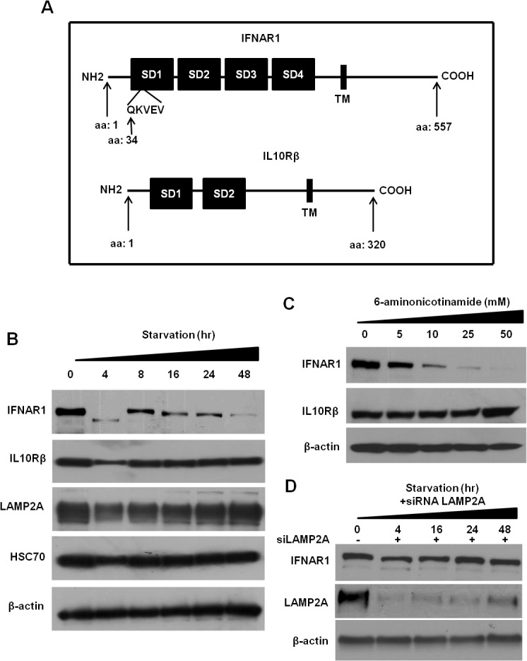
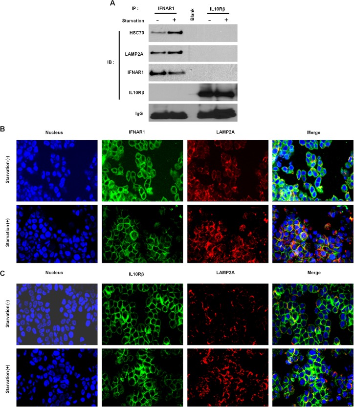
Results: FFA treatment induced dose-dependent hepatocellular steatosis and lipid droplet accumulation in HCV-infected Huh-7.5 cells. FFA treatment of infected culture increased HCV replication in a concentration-dependent manner. Intracellular lipid accumulation led to reduced Stat phosphorylation and nuclear translocation, causing an impaired IFN-α antiviral response and HCV clearance. Type III IFN (IFN-λ), which binds to a separate receptor, induces Stat phosphorylation, and nuclear translocation as well as antiviral clearance in FFA-treated HCV cell culture. We show here that the HCV-induced autophagy response is increased in FFA-treated cell culture. Pharmacological inhibitors of lysosomal degradation, such as ammonium chloride and bafilomycin, prevented IFNAR1 degradation in FFA-treated HCV cell culture. Activators of chaperone-mediated autophagy, including 6-aminonicotinamide and nutrient starvation, decreased IFNAR1 levels in Huh-7.5 cells. Co-immunoprecipitation, colocalization and siRNA knockdown experiments revealed that IFNAR1 but not IFNLR1 interacts with HSC70 and LAMP2A, which are core components of chaperone-mediated autophagy (CMA).

Conclusion: Our study presents evidence indicating that chaperone-mediated autophagy targets IFNAR1 degradation in the lysosome in FFA-treated HCV cell culture. These results provide a mechanism for why HCV induced autophagy response selectively degrades type I but not the type III IFNAR1.


Conflict of interest statement

Competing Interests: The authors have declared that no competing interests exist.

Figures

Fig 1
Fig 1. HCV replication induces microvesicular steatosis in persistently infected Huh-7.5.
(A). Confocal microscopy showing increased accumulation of lipid droplets in Huh-7.5 cells with or without HCV infection. Red: Oil-red staining and green: HCV core expression. The images were taken at 40X magnification. (B). Microfluorometer analysis of intracellular fat accumulation in Huh-7 cells with or without HCV replication. The OD values are compared between uninfected and HCV infected cells. (C). Electron micrograph showing more intracellular lipid droplet accumulation in persistently HCV infected as compared to uninfected Huh-7.5 cell. (D). FFA treatment induces macrovesicular steatosis in Huh-7.5 cells. Uninfected Huh-7.5 cells were treated with increasing concentration of Oleate/Palmitate (2:1 ratio) for 24 hours and hepatocellular steatosis was examined by confocal microscopy after Oil-red staining. (E). Microfluorometer analysis shows dose-dependent intracellular fat accumulation in Huh-7.5 cells treated with oleate and palmitate (FFA). OD values are compared between untreated and treated cells. F. FFA treatment support HCV replication in cell culture. Huh-7.5 cells were infected with JFH1-ΔV3-Rluc and then treated with FFA 0.5mM. Replication of HCV was measured by Renilla luciferase activity.
Fig 2
Fig 2. Persistently infected Huh-7.5 cells were treated with 1xIC90, 2.5 × IC90, and 5 × IC90 IFN-α or IFN-λ.
After 72 hours antiviral efficacy of IFN-α and IFN-λ was determined by the measurement of Renilla luciferase activity and immunohistochemistry for HCV Core protein expression. (A). Normalized Renilla luciferase activity of culture treated with equivalent concentrations of IFN-alpha or IFN-λ. (B). Representative picture showing the expression of HCV core protein in the IFN-treated cells and control uninfected Huh-7.5 by immunostaining. (C). Quantification of HCV Core+ cells in 10 different high-power fields (×40), compared with untreated control. Both assays confirmed that the antiviral effect of IFN-λ is significantly stronger than IFN-α when used at equivalent concentrations (* p< 0.03, *** p< 0.001).
Fig 3
Fig 3. FFA treatment blocks the antiviral response of IFN-α and IFN-γ against HCV.
Infected Huh-7.5 cells were cultured in the presence of FFA for 72 hours and then treated with IFN for additional 72 hours. The relative antiviral effect of IFN-α, IFN-γand IFN-λ was compared in the presence and absence of FFA by measuring Renilla luciferase activity. The luciferase values are normalized with total protein.
Fig 4
Fig 4. Long-term antiviral response of IFN-α, IFN-γ, and IFN-λ against HCV culture with or without FFA treatment.
HCV infected culture was continuously treated with each IFN and antiviral activity was determined by measuring Renilla luciferase activity of cell lysates. (A). Represents normalized Renilla luciferase values of HCV infected culture with or without interferon treatment. (B). Shows the expression of HCV core protein in the IFN-α, IFN-γ, and IFN-λ treated cultures at day 6 and day 20. (C). Represents normalized luciferase values of FFA treated HCV infected culture with or without interferon. (D). Expression of core protein in the in the FFA treated HCV culture after IFN-α, IFN-γ, and IFN-λ treatment at day 6 and day 20.
Fig 5
Fig 5. Persistent HCV infected cell culture with or without FFA treatment impairs IFN-α induced phosphorylation and nuclear translocation of Stat1, Stat2.
(A). Uninfected (HCV-), infected Huh-7.5 cells (HCV+) or infected plus FFA treated cells were treated with IFN-α or IFN-λ or IFN-λ2 for 30 minutes. Equal protein amounts were separated on SDS-PAGE gel and Western blotting was performed using antibodies to p-Stat1, Stat, pStat2 and Stat2 and beta-actin. (B). Nuclear translocation of pSTAT1-GFP and pSTAT2-GFP in the uninfected (left panel) and HCV infected culture (right panel) treated with IFN-α or IFN-λ1. HCV core was detected by immunofluorescense. Red represents HCV core and green represents GFP and blue is nuclear staining.
Fig 6
Fig 6. Jak inhibitor and siRNA targeted to Stat2 prevent IFN-λ antiviral mechanisms against HCV in FFA treated cell culture.
(A and B). FFA treated HCV culture is treated with Jak1 inhibitor for seven days. Antiviral activity of IFN-αand IFN-λwas determined each day by Renilla Luciferase. (C). FFA treated HCV cell culture was treated with siRNA targeting Stat1, Stat2 and Stat3 and after 24 hours culture was treated with IFN-λ. After 72 hours antiviral effect of was determined by Luciferase assay. Silencing Stat2 only prevent IFN-λantiviral activity against HCV in FFA treated culture.
Fig 7
Fig 7. FFA treatment for 48 hours results in down-regulation of cytosolic and membrane expression IFNAR1 in Huh-7.5 cells.
(A). Western blot showing downregulation of cytosolic low molecular weight IFNAR1 as well as mature high molecular weight IFNAR1 in Huh-7.5 cells treated with FFA in a concentration dependent manner. The expression of IFN-λ1 and IFN-λ receptor was not altered. (B). Expression of cytosolic as well as membrane IFNAR1, IFNGR and IFNLRs in FFA treated Huh-7.5 cells. (C). The surface expression of IFNAR1, IFNGR and IFNLR1 in FFA treated Huh-7.5 cells by confocal microscopy.
Fig 8
Fig 8. FFA treatment for 48 hours induced autophagy response in Huh-7.5 cells.
Huh-7.5 cells were treated with increasing concentrations of FFA and after 72 hours and examined for autophagy induction by immunostaining and Western blot analysis. (A) Immunostaining showing expression of p62 in FFA treated Huh-7.5 cells. (B). Western blot show that p62 levels are decreased in FFA treated cells. ATG5 level are induced in FFA treated Huh-7.5 cells. LAMP2A and HSC70 levels and b-actin levels were not altered significantly as compared to untreated Huh-7.5 cells. (C). The expression of p62 was decreased by both HCV and FFA treated culture. The expression of ATG5 and LAMP2A was induced by HCV infected and FFA treated Huh-7.5 cells.
Fig 9
Fig 9. FFA treatment for 48 hours induced decrease of IFNAR1 in Huh-7.5 cells is mediated by a lysosomal degradation pathway.
Huh-7.5 cells were treated with FFA for 24 hours in the presence or absence of lysosome inhibitors such as: NH4Cl (30mM) or BafilomycinA1. The expression levels IFNAR1, IL10Rβ and β-actin were examined using the cell lysates by Western blotting.
Fig 10
Fig 10. Chaperone induced autophagy targets the degradation of IFNAR1 in HCV and FFA treated Huh-7.5 cells for 48 hours.
(A). Schematic representation of IFNAR1 protein where presence of CMA motif site is marked. (B). Uninfected Huh-7.5 cells were cultured in a serum free medium for indicated time points. The expression of IFNAR1 and IFNLR1 as well as CMA proteins (LAMP2A and HSC70) levels were measured by Western blotting. (C). Show the dose-dependent reduced expression of IFNAR1 in the Huh-7.5 cells treated with 6-aminonicotinamide (mM) by Western blotting. The expression of IFN-λ receptor did not change by similar treatment. (D). Measurement of IFNAR1 expression after silencing LAMP2A. Huh-7.5 cells were transfected with 60pmole of siRNA using lipofectamine. After 24 hours, cells were serum starved for 4 to 48 hours and cell lysates were measured for IFNAR1 expression by Western blot analysis.
Fig 11
Fig 11. IFNAR1 binds to LAMP2A and localizes to CMA-associated lysosomes.
(A). Co-immunoprecipitation assay of Huh-7.5 cells serum starved for six hours, after which cells were lysed, and lysates were immunoprecipitated with either IFN-λ receptor or IFN-α receptor and probed for LAMP2A or HSC70 by Western blot analysis. (B). Effect of CMA on IFNAR1 localization with LAMP2A. Confocal images of cellular localization of IFNAR1 (green) and LAMP2A (red) in Huh-7.5 cells cultured with serum free medium. DAPI was used for nuclear stain. Only serum starvation enhances CMA, which is associated with increase signal co-localization (red+green = yellow) indicating that IFNAR1 localizing to CMA associated lysosomes for degradation. (C) Effect of CMA on IFN-λ receptor localization with LAMP2A. There was no increase in signal co-localization.

Similar articles

Cited by

References

    1. Lavanchy D. The global burden of hepatitis C. Liver Int 2009; 29 Suppl 1: 74–81. 10.1111/j.1478-3231.2008.01934.x - DOI - PubMed
    1. Shepard CW, Finelli L, Alter MJ. Global epidemiology of hepatitis C virus infection. Lancet Infect Dis. 2006; 5:558–567. - PubMed
    1. Ghany MG, Nelson DR, Strader DB, Thomas DL, Seeff LB; American Association for Study of Liver Diseases. Hepatology. 2011, 54(4): 1433–44 10.1002/hep.24641 - DOI - PMC - PubMed
    1. Aronsohn A, Jensen D. Interferon-combination strategies for the treatment of chronic hepatitis C. Semin Liver Dis. 2014; 34:30–36. 10.1055/s-0034-1371008 - DOI - PubMed
    1. Forestier N, Zeuzem S. Triple therapy with telaprevir: results in hepatitis C virus-genotype 1 infected relapsers and non-responders. Liver Int. 2012; 32:44–50. 10.1111/j.1478-3231.2011.02720.x - DOI - PubMed

Publication types

MeSH terms

LinkOut - more resources