. 2015 Jun;22(6):476-484.
doi: 10.1038/nsmb.3018. Epub 2015 May 11.

Cryptochrome 1 regulates the circadian clock through dynamic interactions with the BMAL1 C terminus

Affiliations

Cryptochrome 1 regulates the circadian clock through dynamic interactions with the BMAL1 C terminus

Haiyan Xu et al. Nat Struct Mol Biol. 2015 Jun.

Abstract

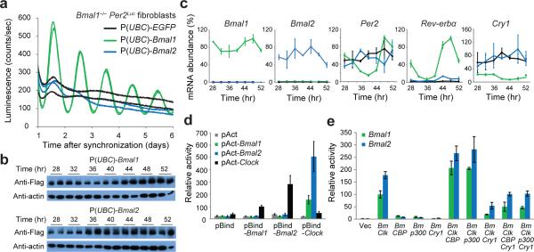
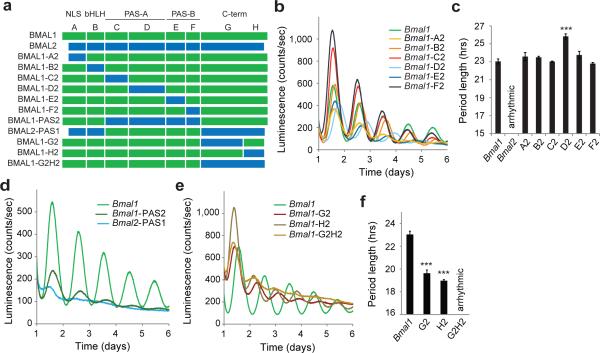
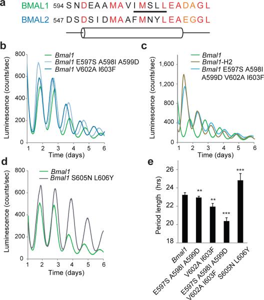
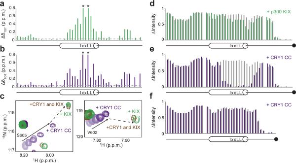
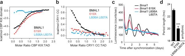
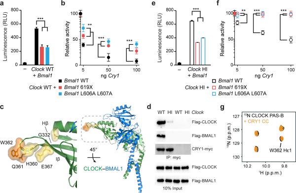
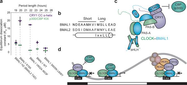
The molecular circadian clock in mammals is generated from transcriptional activation by the bHLH-PAS transcription factor CLOCK-BMAL1 and subsequent repression by PERIOD and CRYPTOCHROME (CRY). The mechanism by which CRYs repress CLOCK-BMAL1 to close the negative feedback loop and generate 24-h timing is not known. Here we show that, in mouse fibroblasts, CRY1 competes for binding with coactivators to the intrinsically unstructured C-terminal transactivation domain (TAD) of BMAL1 to establish a functional switch between activation and repression of CLOCK-BMAL1. TAD mutations that alter affinities for co-regulators affect the balance of repression and activation to consequently change the intrinsic circadian period or eliminate cycling altogether. Our results suggest that CRY1 fulfills its role as an essential circadian repressor by sequestering the TAD from coactivators, and they highlight regulation of the BMAL1 TAD as a critical mechanism for establishing circadian timing.


Figures

Figure 1
Figure 1
Only BMAL1 can restore circadian rhythms in Bmal1–/– Per2Luc fibroblasts. (a) Luminescence records from Bmal1–/– Per2Luc fibroblasts expressing EGFP, Bmal1 or Bmal2. Traces from replicate cell cultures are shown from one representative experiment (n = 3 clonal lines). (b) Western blot of Flag-tagged Bmal paralog expression in Bmal1–/– Per2Luc fibroblasts. Time, hours after synchronization. Uncropped images can be found in Supplementary Data Set 1. (c) Expression of clock-controlled genes in Bmal1–/– Per2Luc fibroblasts determined by quantitative reverse transcription PCR. Values are expressed as percentage of maximum expression, set to 100% for each gene except for Bmal2, which was normalized to Bmal1 based on PCR amplification efficiency to reflect their relative levels. Points represent mean expression ± s.d. of triplicate measurements from two independent timecourses. Time, hours after synchronization. (d) Luciferase-based mammalian two-hybrid assay in HEK293T cells transiently transfected with the indicated plasmid pairs. pBind, fusion with Gal4 DNA-binding domain; pAct, fusion with VP16 transactivation domain. Luminescence values are expressed as mean ± s.d. of triplicate measurements from one representative experiment (n = 3). Relative activity normalized to pAct-Clock–pBind-Bmal1 set to 100. (e) Per1-Luc assay of CLOCK–BMAL activity in HEK293T cells transiently transfected with indicated plasmids. Vec, vector; Bm, Bmal1 or Bmal2; Clk, Clock. Luminescence values are expressed as mean ± s.d. of triplicate measurements from one representative experiment (n = 3). Relative activity normalized to ClockBmal1 set to 100.
Figure 2
Figure 2
The BMAL1 C-terminus is needed for circadian function. (a) Domain organization of BMAL proteins and diagram of chimeric constructs. Chimera boundaries can be found in Supplemental Figure 2. (b) Luminescence records from Bmal1–/– Per2Luc fibroblasts expressing WT Bmal1 or Bmal1 chimeras with Bmal2 substitutions of the N-terminal core domains. One trace per chimera is shown from a representative experiment (n = 3 clonal lines with 2 cell culture replicates each). (c) Mean period of rescued circadian luminescence rhythms ± s.d. (n = 3 clonal lines with 2 cell culture replicates each). (d) Luminescence records from Bmal1–/– Per2Luc fibroblasts expressing WT Bmal1 or PAS-swapping chimeras. One trace per chimera is shown from a representative experiment (n = 3 clonal lines with 2 cell culture replicates each). (e) Luminescence records from Bmal1–/– Per2Luc fibroblasts expressing WT Bmal1 or C-terminal chimeras. One trace per chimera is shown from a representative experiment (n = 3 clonal lines with 2 cell culture replicates each). (f) Mean period of rescued circadian luminescence rhythms ± s.d. (n = 3 clonal lines with 2 cell culture replicates each). *** P < 0.001 compared to WT Bmal1, two-tailed paired t test.
Figure 3
Figure 3
A helical motif within the BMAL1 H domain controls circadian period length. (a) Comparison of BMAL paralog sequence and predicted secondary structure near the H domain α-helix. Underlined, KIX-binding IxxLL motif. Residue coloring: red, conserved; orange, similar; black, non-conserved. Cylinder, predicted α-helix. (b-d) Luminescence records from Bmal1–/– Per2Luc fibroblasts expressing WT Bmal1 or Bmal1 chimeras with substitutions from the Bmal2 H domain α-helix, including mutants of the N-terminal (b-c) or C-terminal half of the α-helix (d). One trace per chimera is shown from a representative experiment (n = 3 clonal lines with 2 cell culture replicates each). (e) Mean period of rescued circadian luminescence rhythms ± s.d. (n = 3 clonal lines with 2 cell culture replicates each). ** P < 0.01, *** P < 0.001 compared to WT Bmal1, two-tailed paired t test.
Figure 4
Figure 4
CBP(p300) and CRY1 interact with BMAL1 TAD. (a-b) Backbone chemical shift perturbations of 15N BMAL1 TAD with stoichiometric p300 KIX (a) or CRY1 CC (b). p.p.m., parts per million. Dashed line, ΔδTOT significance cutoff = 0.04 p.p.m. *residues broadened beyond detection. (c) Highlighted regions of 15N HSQC spectra showing titration of 15N BMAL1 TAD with CRY1 CC to 1:1 stoichiometry (light to dark purple; solid arrow); at 1:1 stoichiometry with p300 KIX (green); or in the presence of both CRY1 CC and p300 KIX (brown). Dashed line shows transition of 15N TAD–CRY1 to 15N TAD–p300 KIX upon competition. (d-e) Relative15N TAD–EDTA peak intensities from PRE experiments as ratio of Mn2+ chelation by the C-terminal EDTA (black circle) in the free protein (gray), or upon complex formation with p300 KIX (green) (d) or CRY1 CC (purple) (e). (f) Relative15N TAD 619X–EDTA peak intensities from PRE experiments as ratio of Mn2+ chelation by the C-terminal EDTA (black circle) in the free protein (gray), or upon complex formation with CRY1 CC (purple).
Figure 5
Figure 5
BMAL1 TAD mutations decrease affinity for CBP(p300) and CRY1 and influence circadian period. (a) Isothermal titration calorimetric (ITC) profiles for the interaction of CBP KIX domain with WT or mutant BMAL1 TADs. Line, one-site binding model representing best fit to data; see Table 1. (b) ITC profile for the interaction of CRY1 CC peptide with WT or mutant BMAL1 TADs. (c) Luminescence records from Bmal1–/– Per2Luc fibroblasts expressing WT or mutant Bmal1. One trace per chimera is shown from a representative experiment (n = 3 clonal lines with 2 cell culture replicates each). (d) Mean period of rescued circadian luminescence rhythms ± s.d. (n = 3 clonal lines with 2 cell culture replicates each). *** P < 0.001 compared to WT Bmal1, two-tailed paired t test.
Figure 6
Figure 6
Distinct sites on CLOCK and BMAL1 facilitate repression by CRY1. (a) Per1-Luc assay in HEK293T cells transiently transfected with plasmids encoding Clock WT and Bmal1 WT, 619X or L606A L607A. Relative light units (RLU) are scaled down by 103 and expressed as mean ± s.d. of triplicate measurements from one representative experiment (n = 3). (b) Per1-Luc assay with increasing amounts of plasmids encoding Cry1 with Clock WT and Bmal1 WT, 619X or L606A L607A. Relative activity normalized to Clock WT–Bmal1 WT set to 100. (c) Residues in the Hβ-Iβ (HI) loop on CLOCK PAS-B (PDB 4F3L) that reduce CRY1 repression when mutated (yellow or orange). (d) Coimmunoprecipitation (IP) of Flag-CLOCK WT or HI (Q361P W362R) and Flag-BMAL1 by CRY1-myc from HEK293T cells with anti-myc antibody. Western blots (IB) were performed using indicated antibodies. Uncropped images can be found in Supplementary Data Set 1. (e) Per1-Luc assay with plasmids encoding Clock WT and Bmal1 WT, 619X or L606A L607A. RLU represented as in panel (a). (f) Per1-Luc assay with increasing amounts of plasmid encoding Cry1 with Clock HI and Bmal1 WT, 619X or L606A L607A. Relative activity normalized to Clock HI–Bmal1 WT set to 100. (g) Tryptophan indole region of N HSQC spectra showing 15N CLOCK PAS-B in the absence (black) and presence (orange) of CRY1 CC. The full spectrum can be found in Supplementary Fig. 8. ** P < 0.01; ***P < 0.001 compared to Bmal1 WT by two-tailed paired t test.
Figure 7
Figure 7
Regulation of the BMAL1 TAD by CRY1 contributes to determination of circadian period. (a) Correlation of coregulator affinities for isolated TADs with period lengths from circadian rescue experiments. Not shown: Bmal1 L606A L607A mutant, which has no observable affinity for either CBP(p300) KIX or CRY1 CC and does not rescue circadian rhythms in Bmal1–/– Per2Luc fibroblasts. (b) Location of long and short period substitution mutants in the TAD α-helix. (c) Model of CRY1 interactions with the CLOCK–BMAL1 complex. (d) Model for regulation of the circadian transcriptional feedback loop by the ternary CLOCK–BMAL1–CRY1 complex. Black and white bars underneath indicate night or day, respectively. See text for more details.

Comment in

References

    1. Bass J. Circadian topology of metabolism. Nature. 2012;491:348–356. - PubMed
    1. Partch CL, Green CB, Takahashi JS. Molecular architecture of the mammalian circadian clock. Trends in cell biology. 2014;24:90–99. - PMC - PubMed
    1. Brown S, et al. PERIOD1-associated proteins modulate the negative limb of the mammalian circadian oscillator. Science. 2005;308:693–696. - PubMed
    1. Duong H, Robles M, Knutti D, Weitz C. A molecular mechanism for circadian clock negative feedback. Science. 2011;332:1436–1439. - PMC - PubMed
    1. Lee C, Etchegaray J, Cagampang F, Loudon A, Reppert S. Posttranslational mechanisms regulate the mammalian circadian clock. Cell. 2001;107:855–867. - PubMed

Publication types