

Cognitive Improvement after Mild Traumatic Brain Injury Measured with Functional Neuroimaging during the Acute Period
- PMID: 25962067
- PMCID: PMC4427352
- DOI: 10.1371/journal.pone.0126110
Cognitive Improvement after Mild Traumatic Brain Injury Measured with Functional Neuroimaging during the Acute Period
Abstract
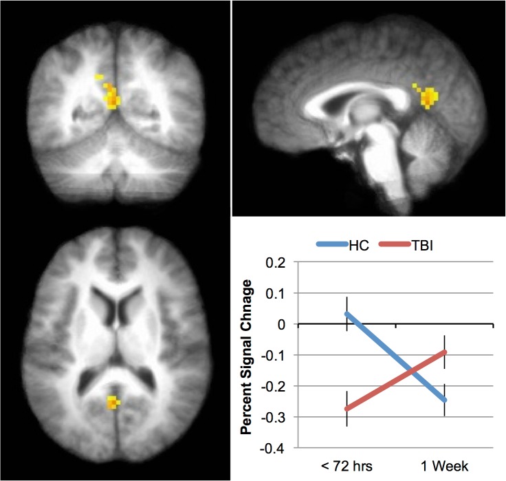
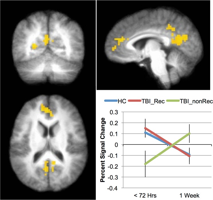
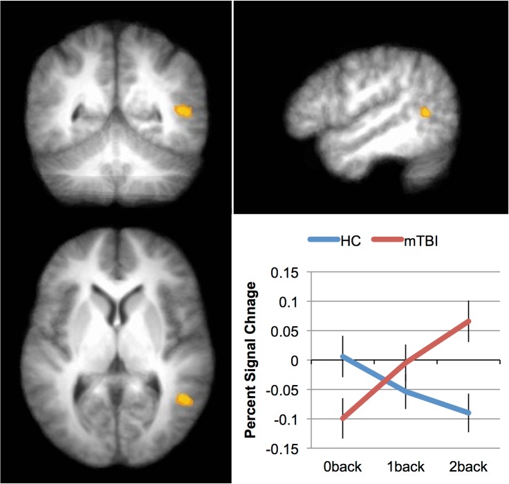
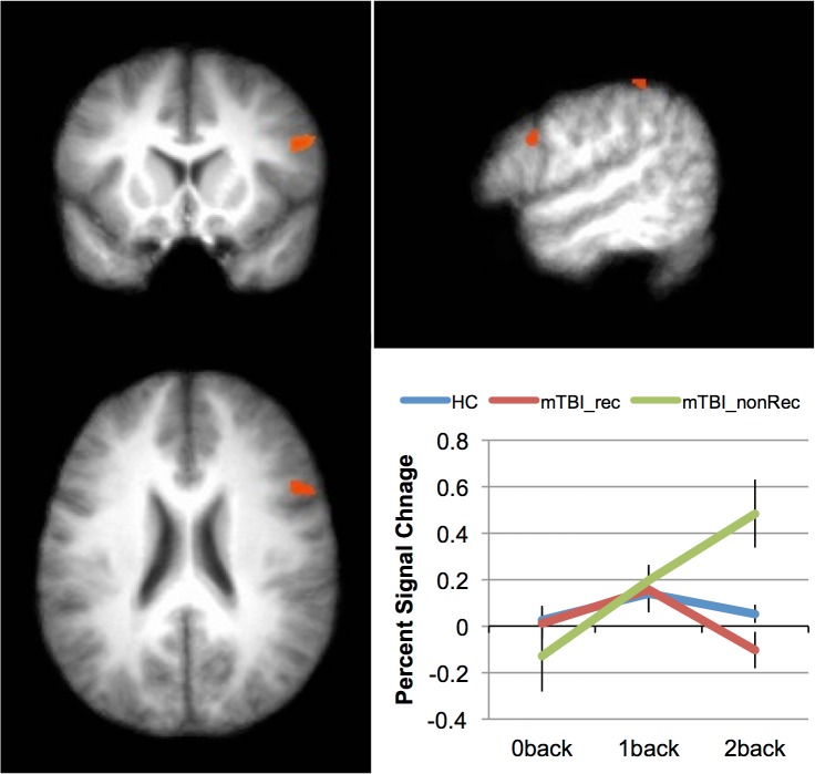
Functional neuroimaging studies in mild traumatic brain injury (mTBI) have been largely limited to patients with persistent post-concussive symptoms, utilizing images obtained months to years after the actual head trauma. We sought to distinguish acute and delayed effects of mild traumatic brain injury on working memory functional brain activation patterns < 72 hours after mild traumatic brain injury (mTBI) and again one-week later. We hypothesized that clinical and fMRI measures of working memory would be abnormal in symptomatic mTBI patients assessed < 72 hours after injury, with most patients showing clinical recovery (i.e., improvement in these measures) within 1 week after the initial assessment. We also hypothesized that increased memory workload at 1 week following injury would expose different cortical activation patterns in mTBI patients with persistent post-concussive symptoms, compared to those with full clinical recovery. We performed a prospective, cohort study of working memory in emergency department patients with isolated head injury and clinical diagnosis of concussion, compared to control subjects (both uninjured volunteers and emergency department patients with extremity injuries and no head trauma). The primary outcome of cognitive recovery was defined as resolution of reported cognitive impairment and quantified by scoring the subject's reported cognitive post-concussive symptoms at 1 week. Secondary outcomes included additional post-concussive symptoms and neurocognitive testing results. We enrolled 46 subjects: 27 with mild TBI and 19 controls. The time of initial neuroimaging was 48 (+22 S.D.) hours after injury (time 1). At follow up (8.7, + 1.2 S.D., days after injury, time 2), 18 of mTBI subjects (64%) reported moderate to complete cognitive recovery, 8 of whom fully recovered between initial and follow-up imaging. fMRI changes from time 1 to time 2 showed an increase in posterior cingulate activation in the mTBI subjects compared to controls. Increases in activation were greater in those mTBI subjects without cognitive recovery. As workload increased in mTBI subjects, activation increased in cortical regions in the right hemisphere. In summary, we found neuroimaging evidence for working memory deficits during the first week following mild traumatic brain injury. Subjects with persistent cognitive symptoms after mTBI had increased requirement for posterior cingulate activation to complete memory tasks at 1 week following a brain injury. These results provide insight into functional activation patterns during initial recovery from mTBI and expose the regional activation networks that may be involved in working memory deficits.
Conflict of interest statement
Figures
References
-
- Faul MX L, Wald MM, Coronado VG. Traumatic Brain Injury in the United States: Emergency Department Visits, Hospitalizations and Deaths 2002–2006. Centers for Disease Control and Prevention, National Center for Injury Prevention and Control. 2010;Atlanta (GA).
Publication types
MeSH terms
Grants and funding
LinkOut - more resources
Full Text Sources
Other Literature Sources
Medical
Research Materials
