. 2015 May 11;10(5):e0125690.
doi: 10.1371/journal.pone.0125690. eCollection 2015.

Cellular promyelocytic leukemia protein is an important dengue virus restriction factor

Affiliations

Cellular promyelocytic leukemia protein is an important dengue virus restriction factor

Federico Giovannoni et al. PLoS One. .

Erratum in

Abstract

The intrinsic antiviral defense is based on cellular restriction factors that are constitutively expressed and, thus, active even before a pathogen enters the cell. The promyelocytic leukemia (PML) nuclear bodies (NBs) are discrete nuclear foci that contain several cellular proteins involved in intrinsic antiviral responses against a number of viruses. Accumulating reports have shown the importance of PML as a DNA virus restriction factor and how these pathogens evade this antiviral activity. However, very little information is available regarding the antiviral role of PML against RNA viruses. Dengue virus (DENV) is an RNA emerging mosquito-borne human pathogen affecting millions of individuals each year by causing severe and potentially fatal syndromes. Since no licensed antiviral drug against DENV infection is currently available, it is of great importance to understand the factors mediating intrinsic immunity that may lead to the development of new pharmacological agents that can boost their potency and thereby lead to treatments for this viral disease. In the present study, we investigated the in vitro antiviral role of PML in DENV-2 A549 infected cells.


Conflict of interest statement

Competing Interests: The authors have declared that no competing interests exist.

Figures

Fig 1
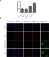
Fig 1. Assessment of PML silencing and overexpression in A549 cells.
A549 cells were non-transfected, transfected with X-siRNAs, PML-siRNAs or pcDNA-PMLIV. (A) At 24 h post transfection, cells were fixed and PML protein was stained using anti-PML monoclonal antibody and TRITC-labeled anti-mouse IgG. Cells were visualized by fluorescence microscopy. Magnification: 400 X. (B) In parallel, cells were harvested for determination of PML-mRNA expression levels by real time PCR. PML-mRNA expression level is represented as fold difference relative to X-siRNAs-transfected cells and normalized to β-actin-mRNA. The reported values are mean ± SD (n = 3). Asterisks indicate a significant difference (*** p < 0.001; ** p < 0.01; * p < 0.05). (C) Counts of apoptotic cells and apoptotic bodies were performed using 400 X magnification. All identified apoptosis in the sample were counted and the % of apoptotic cells was defined as the total number of apoptotic cells and apoptotic bodies in at least 500 cells.
Fig 2
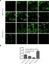
Fig 2. Effect of PML silencing and overexpression on infectious particle production and antigen expression.
A549 cells were non-transfected, transfected with X-siRNAs, PML-siRNAs or pcDNA-PMLIV and infected with DENV-2. Non-infected cells were also included as a control (A) At 24 h p.i. viral yields were determined by a standard plaque assay. The reported values are mean ± SD (n = 3). Asterisks indicate a significant difference (*** p < 0.001; ** p < 0.01; * p < 0.05). (B) In parallel, cells were fixed at 24 h p.i. and viral glycoprotein E was stained using anti-E monoclonal antibody and FITC-labeled anti-mouse IgG2a. Cells were visualized by fluorescence microscopy. Magnification: 400 X. (C) Quantification of DENV-2 antigen expressing cells shown in B.
Fig 3
Fig 3. Effect of DENV-2 infection on PML-NBs distribution, number and size.
DENV-2 infected A549 cells were fixed 24 h p.i. and a double immunofluorescence staining was performed. (A) Viral antigen expression was visualized by using monoclonal anti-E antibody and FITC-labelled anti-mouse IgG2a. PML was visualized by using anti-PML polyclonal antibodies and TRITC-labelled anti-rabbit IgG1. Cell nuclei were stained with 4',6-diamidino-2-phenylindole (DAPI). Cells were visualized by confocal microscopy. Magnification: 600 X. (B) Quantification of the mean number of PML-NBs in the nuclei of non-infected, DENV-2 infected and DENV-2 infected neighboring A549 cells at 24 h p.i. 50 cell nuclei were counted for each condition. The reported values are mean ± SD. (C) Determination of the mean diameter of PML-NBs in non-infected, DENV-2 infected and DENV-2 infected neighboring A549 cells represented as a box and whiskers plot. The box extends from the 25th percentile to the 75th percentile. The horizontal line represents the median and the whiskers extend down to the smallest value and up to the largest. PML-NB sizes from 100 non-infected, DENV-2 infected or DENV-2 infected neighboring cells were measured. Asterisks indicate a significant difference (*** p < 0.001; ** p < 0.01; * p < 0.05).
Fig 4
Fig 4. Determination of PML expression with the time of infection.
(A) A549 cells were infected with DENV-2 (MOI = 1) and at 2, 4, 16 and 24 h p.i. the expression of PML-mRNA was determined by qRT-PCR. (B) A549 cells were infected with DENV-2 (MOI = 1) and at 2, 4, 18 and 48 h p.i. the expression of PML was revealed by immunofluorescence with anti-PML monoclonal antibodies. Magnification: 400X.
Fig 5
Fig 5. Determination of PML localization during infection.
(A) A549 cells were infected with DENV-2 (MOI = 1) and at 24 and 48 h p.i. the expression of PML was revealed by confocal microscopy with anti-PML polyclonal antibodies. Both results are shown in consecutive confocal partial z-stack images. (B) Quantification of cellular PML expression level shown in C.
Fig 6
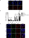
Fig 6. Effect of IFN-I on PML anti-DENV-2 activity.
(A) A549 cells were treated with fresh medium (FM), conditioned medium (CM) and medium containing 2500 U/ml of IFN-I during 24 h. Then, cells were fixed and immunostained with anti-PML monoclonal antibodies. (B) A549 and Vero cells were infected with DENV-2 (MOI = 1) and 24 h p.i. the expression of TLR3, RIG-I, TRAF6, IL-6, IFN-b and PML mRNA was determined by real time PCR. A549 cells infected with UV-inactivated DENV-2 (A549-UV) and DENV-2 infected A549 cells treated with 10 μM JAK inhibitor I (A549-JAK inhibitor I) were included for comparison. The mRNA expression level is represented as fold difference relative to mock infected cells and normalized to β-actin-mRNA. (C) Non-infected Vero cells, DENV-2 infected Vero cells and non-infected Vero cells treated with 2500 U/ml of IFN-I (IFN) were incubated during 24 h. Then, cells were fixed and immunostained with anti-PML polyclonal antibodies. Magnification: 600 X.

References

    1. Saffert RT, Kalejta RF (2008) Promyelocytic leukemia-nuclear body proteins: herpesvirus enemies, accomplices, or both? Future Virol 3: 265–277. - PMC - PubMed
    1. Tavalai N, Papior P, Rechter S, Stamminger T (2008) Nuclear domain 10 components promyelocytic leukemia protein and hDaxx independently contribute to an intrinsic antiviral defense against human cytomegalovirus infection. J Virol 82: 126–37. - PMC - PubMed
    1. Geoffroy MC, Chelbi-Alix MK (2011) Role of Promyelocytic Leukemia Protein in Host Antiviral Defense. J Interferon Cytokine Res 31: 145–158. 10.1089/jir.2010.0111 - DOI - PubMed
    1. Borden KL (2002) Pondering the promyelocytic leukemia protein (PML) puzzle: possible functions for PML nuclear bodies. Mol Cell Biol 22: 5259–69. - PMC - PubMed
    1. Maul GG, Negorev D, Bell P, Ishov AM (2000) Review: properties and assembly mechanisms of ND10, PML bodies, or PODs. J Struct Biol 129: 278–87. - PubMed

Publication types