. 2015 May 11;10(5):e0120061.
doi: 10.1371/journal.pone.0120061. eCollection 2015.

Pathogenicity of Highly Pathogenic Avian Influenza Virus H5N1 in Naturally Infected Poultry in Egypt

Affiliations

Pathogenicity of Highly Pathogenic Avian Influenza Virus H5N1 in Naturally Infected Poultry in Egypt

Ibrahim Thabet Hagag et al. PLoS One. .

Abstract

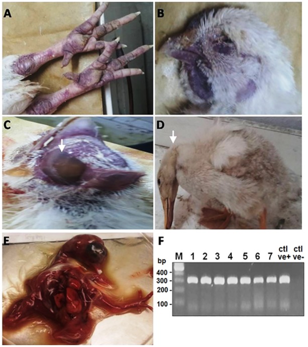
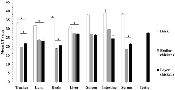
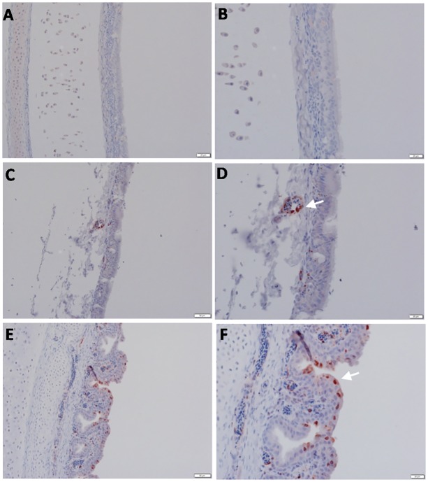
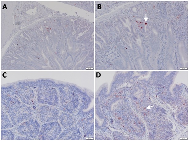
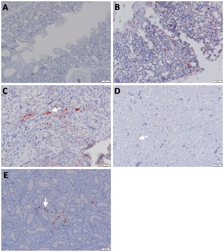
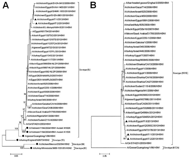
Highly pathogenic avian influenza virus (HPAIV) H5N1 has been endemic in Egypt since 2006, and there is increasing concern for its potential to become highly transmissible among humans. Infection by HPAIV H5N1 has been described in experimentally challenged birds. However, the pathogenicity of the H5N1 isolated in Egypt has never been reported in naturally infected chickens and ducks. Here we report a 2013 outbreak of HPAIV H5N1 in commercial poultry farms and backyards in Sharkia Province, Egypt. The main symptoms were ecchymosis on the shanks and feet, cyanosis of the comb and wattles, subcutaneous edema of the head and neck for chickens, and nervous signs (torticollis) for ducks. Within 48-72 hrs of the onset of illness, the average mortality rates were 22.8-30% and 28.5-40% in vaccinated chickens and non-vaccinated ducks, respectively. Tissue samples of chickens and ducks were collected for analyses with cross-section immunohistochemistry and real-time RT-PCR for specific viral RNA transcripts. While viral RNA was detected in nearly all tissues and sera collected, viral nucleoprotein was detected almost ubiquitously in all tissues, including testis. Interestingly, viral antigen was also observed in endothelial cells of most organs in chickens, and clearly detected in the trachea and brain in particular. Viral nucleoprotein was also detected in mononuclear cells of various organs, especially pulmonary tissue. We performed phylogenetic analyses and compared the genomic sequences of the hemagglutinin (HA) and nonstructural proteins (NS) among the isolated viruses, the HPAIV circulated in Egypt in the past and currently, and some available vaccine strains. Further analysis of deduced amino acids of both HA and NS1 revealed that our isolates carried molecular determinants of HPAIV, including the multibasic amino acids (PQGERRRK/KR*GLF) in the cleavage site in HA and glutamate at position 92 (D92E) in NS1. This is the first report of the pathogenicity of the HPAIVH5N1 strain currently circulating in naturally infected poultry in Egypt, which may provide unique insights into the viral pathogenesis in HPAIV-infected chickens and ducks.


Conflict of interest statement

Competing Interests: The authors have declared that no competing interests exist.

Figures

Fig 1
Fig 1. Naturally infected chickens and ducks by HPAIV H5N1.
(A) Chickens: subcutaneous hemorrhages in non-feathered skin. (B) Chickens: cyanosis of comb & wattles. (C) Chickens: facial edema. (D) Ducks: nervous signs, torticollis. (E) Infected chicken embryos: chicken embryos were inoculated with supernatants of homogenized testicular tissue of naturally infected layer chickens; the inoculated embryos died within 48hrs with severe congestion and hemorrhages after the first passage. (F) PCR amplification for HA gene showing bands of 317bp in size. First lane: Molecular marker of 100 bp, Lane 1–7: Positive samples, Ctrl +ve: Positive AIV subtype H5N1, Ctrl-ve: Negative control.
Fig 2
Fig 2. Detection of M gene transcripts by RT-PCR.
Results were expressed as Ct values; Ct values lower than (35) were considered positive; Ct values between 15 and 25 were considered strong positive, while those between 25 and 30 were considered moderate positive; Ct values between 30 and 35 were considered weak positive. The tests were performed in triplicates for each sample and the Ct numbers are average with 3xSD (* P<0.05).
Fig 3
Fig 3. Detection of viral NP antigen in trachea by IHC.
(A and B) Trachea of non-infected birds (X = 50 and 20μm, respectively). (C and D) Endothelial cells of trachea of infected birds (X = 50 and 20μm, respectively). (E and F) Epithelial cells of trachea of infected birds (X = 50 and 20μm, respectively).
Fig 4
Fig 4. Detection of viral NP antigen in brain by IHC.
(A and B) Brain of non-infected birds (X = 50 and 20μm, respectively). (C) Perkinji cell layer of cerebellum of infected birds (X = 50μm). (D) Neurons and glial cells of perkinji cell layer of cerebellum of infected birds (arrow) (X = 20μm). (E) Endothelial cells of perkinji cell layer of cerebellum of infected birds (arrow) (X = 20μm).
Fig 5
Fig 5. Detection of viral NP antigen in spleen and pancreas by IHC.
(A and B) Spleen of non-infected birds (X = 50 and 20μm, respectively). (C) Lymphocytes in spleen of infected birds (X = 50μm). (D) Lymphocytes and mononuclear cells in spleen of infected birds (arrow) (X = 20μm). (E and F) Acinar epithelium of pancreas of infected birds (X = 50 and 20μm, respectively).
Fig 6
Fig 6. Detection of viral NP antigen in proventriculus and bursa of Fabricius by IHC.
(A) Proventriculus of infected birds (X = 50μm). (B) Glandular epithelium of proventriculus (arrow) (X = 20μm). (C and D) Lymphocytes in follicular layer of bursa of Fabricius of infected birds (arrow) (X = 50 and 20μm, respectively).
Fig 7
Fig 7. Detection of viral NP antigen in lung, liver and testisby IHC.
(A) Lung of non-infected birds (X = 50μm). (B and C) Macrophages and mononuclear cells in lungs of infected birds (arrow) (X = 50 and 20μm, respectively). (D) Liver sinusoids of infected birds (inside van Kuppfer cells) (arrow) (X = 50μm). (E) Testis of infected birds (between seminephrous tubules of testicular tissue) (arrow) (X = 50μm).
Fig 8
Fig 8. Phylogenetic analysis of nucleotide sequences of the HA and NS genes.
(A) Phylogenetic analysis of HA gene nucleotide sequences at the cleavage site. The sequences of known HPAIV HA genes were obtained from GenBank. Vaccine H5 strains were included in the tree and marked with solid circles. The tree was constructed with multiple alignment of a 309 base-nucleotide sequence of HA genes using the Neighborhood-joining method in MEGA5. The tree topology was evaluated by 1,000 bootstrap analyses. The viruses isolated in this study are marked with solid triangle. (B) Phylogenetic analysis of NS gene on the basis of nucleotide sequences of the complete coding region (822 bases). The tree was constructed using Neighborhood joining method with bootstrap values calculated for 1,000 replicates and cut off value of 50%. Sequences of the isolates from this study are marked with solid triangles.

References

    1. Xu X, Subbarao K, Cox NJ, Guo Y. Genetic characterization of the pathogenic influenza A/Goose/Guangdong/1/96 (H5N1) virus: similarity of its hemagglutinin gene to those of H5N1 viruses from the 1997 outbreaks in Hong Kong. Virology. 1999;261(1):15–9. - PubMed
    1. Sims L, Ellis T, Liu K, Dyrting K, Wong H, Peiris M, et al. Avian influenza in Hong Kong 1997–2002. Avian diseases. 2003;47(s3):832–8. - PubMed
    1. Sims L, Domenech J, Benigno C, Kahn S, Kamata A, Lubroth J, et al. Origin and evolution of highly pathogenic H5N1 avian influenza in Asia. Veterinary Record. 2005;157(6):159 - PubMed
    1. Chen H, Smith G, Zhang S, Qin K, Wang J, Li K, et al. Avian flu: H5N1 virus outbreak in migratory waterfowl. Nature. 2005;436(7048):191–2. - PubMed
    1. Liu J, Xiao H, Lei F, Zhu Q, Qin K, Zhang X-w, et al. Highly pathogenic H5N1 influenza virus infection in migratory birds. Science. 2005;309(5738):1206-. - PubMed

Publication types

MeSH terms

Substances