

doi: 10.1002/adma.201500417.
Epub 2015 May 12.
Enzyme-Responsive Delivery of Multiple Proteins with Spatiotemporal Control
Affiliations
- PMID: 25962336
- PMCID: PMC4633528
- DOI: 10.1002/adma.201500417
Item in Clipboard
Enzyme-Responsive Delivery of Multiple Proteins with Spatiotemporal Control
Adv Mater.
.
Abstract
Orchestrated biological materials such as enzymes and growth factors regulate the growth of tissues and organs. A chirality-controlled, single-protein technology is devised to tailor the spatiotemporally defined delivery of therapeutic proteins in response to natural enzymes present at wound sites. Sustained delivery of one protein and sequential delivery of two proteins are demonstrated for stroke and skin wound healing.
Keywords: chiral peptides; enzyme responsive; protein delivery; sequential release; wound healing.
© 2015 WILEY-VCH Verlag GmbH & Co. KGaA, Weinheim.
Figures
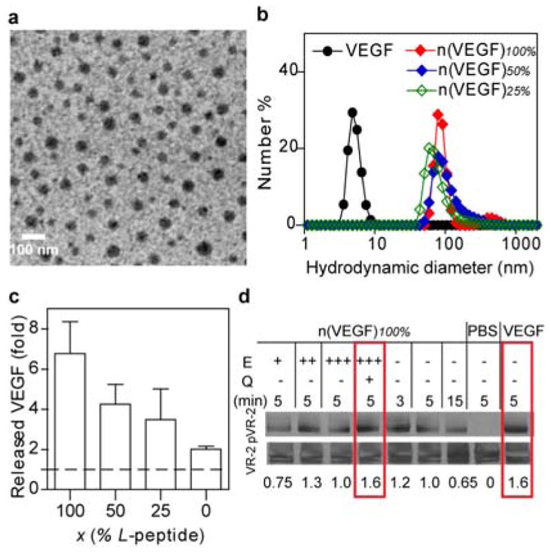
Characterizations of protein nanocapsules. (a) A transmission electron microscopic image of nanocapsules synthesized with gold nanoparticle (AuNP, 4 nm)-labeled VEGF. AuNP is stained with silver enhancer kit to exhibit as dark black dots within the grey spheres of n(VEGF-AuNP)100%. (Scale bar 100 nm) (b) The hydrodynamic sizes of n(VEGF) of different L-to-D ratios via dynamic light scattering. (c) Incubating n(VEGF)100%, n(VEGF)50%, n(VEGF)25% or n(VEGF)0% with plasmin for 20 minutes in ELISA measures the in vitro enzymatic release rates of nanocapsules. (d) Western blotting analysis of the activity of encapsulated VEGF remains identical to free VEGF in inducing receptor phosphorylation. The encapsulated VEGF was first released from n(VEGF)100% at 50 ng/mL via incubation with an enzyme (E), trypsin, at increasing mass ratios of trypsin:n(VEGF)100% = 0.05:1 (labeled as E +), 0.1:1 (labeled as E ++), and 0.2:1 (labeled as E+++). To prevent the released VEGF from being degraded, an inhibitor, aprotinin, was subsequently used to quench (Q) the excessive proteolytic activity of trypsin. Released VEGF, as well as n(VEGF)100% without enzyme pretreatment and free VEGF (50 ng/mL) were incubated with serum-starved human vein endothelial cells for specified amounts of time (min). The activity was indicated by the normalized intensity amounts with the phosphorylation of VEGF receptor-2 normalized by actin.
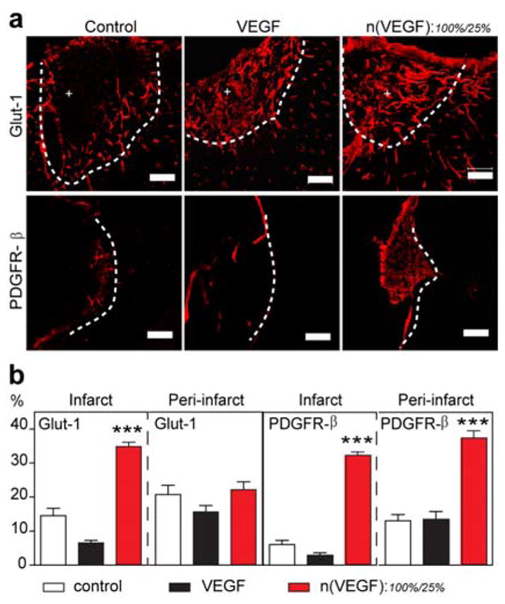
Temporal control of VEGF delivery in mouse stroke model. (a) Representative confocal images of blood vessels (Glut-1+) and their maturity markers (PDGFRβ+) in the infarct (indicated by +) and peri-infarct areas of stroke (separated by the dashed line). The stroke was treated with in situ crosslinked, adhesion peptide-modified hyaluronic acid hydrogels containing no VEGF (control), unencapsulated VEGF (200 ng), or n(VEGF)100% : n(VEGF)25% at 100 ng : 100 ng. (b) Analysis of Glut-1 and PDGFR-β markers for the vascularization in the infarct and peri-infarct areas of stroke. (AVONA with Tukey’s post test, mean ± SEM, N = 3~4, * p<0.05, ** p<0.01, *** p<0.001.) (Scale bar, 100 μm).
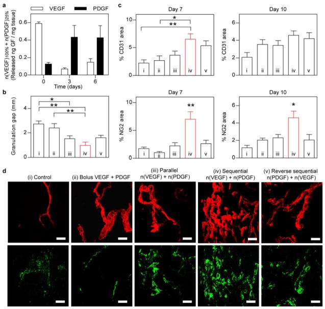
In vivo co-delivery of VEGF and PDGF with temporal control in diabetic mouse skin wounds. (a) Tissue-ELISA analysis of wound-mediated release of proteins from n(VEGF)100% and n(PDGF)25% at day 0, 3, and 6 post surgery. (b) Quantification of the gap of granulation tissues in diabetic skin wounds at day 7. Wound was dressed with fibrin matrices that contained (i) no growth factors (control), (ii) un-encapsulated VEGF (200 ng) and PDGF (200 ng), (iii) mixture of n(VEGF)100%/n(VEGF)25%/n(PDGF)100%/n(PDGF)25% at 100 ng/100 ng/100 ng/100 ng (parallel), (iv) mixture of n(VEGF)100%/n(VEGF)25%/n(PDGF)25%/n(PDGF)10% at 100 ng/100 ng/100 ng/100 ng (sequential), and (v) mixture of n(VEGF)25%/n(VEGF)10%/n(PDGF)100%/n(PDGF)25% at 100 ng/100 ng/100 ng/100 ng (reverse sequential). (c) Immunohistochemical analysis of vessel endothelium (CD31+) and pericyte coverage (NG2+) at day 7 and day 10. (d) Representative confocal images of CD31 (upper row) and NG2 (lower row) in the granulation tissue of diabetic skin wounds at day 7. (AVONA with Tukey’s post test, mean ± SEM, N = 4~6, * p<0.05, ** p<0.01.) Scale bar = 50 μm (d).
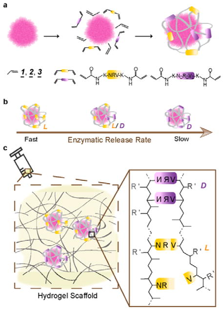
Illustration of chirality-controlled, enzyme-responsive protein nanocapsules with temporal control. (a). The synthesis of the nanocapsules by enriching monomers and crosslinkers around an individual protein molecule and by subsequent in situ polymerization. The monomers can be acrylamide (1 , neutral), N-(3-aminopropyl)methacrylamide (2 , positively charged), or 2-acrylamino-2-methyl-1-propanesulfonic acid (3 , negatively charged). The crosslinkers include the mixtures with designed molar ratios of L (yellow) and D (purple) enantiomers of the peptide Asn-Arg-Val, being the substrate of plasmin. (b) The rate of enzymatic degradations of individual nanocapsule is tuned by varying the ratio of L to D peptide crosslinkers used: faster with more L peptide, slower with more D peptide. (c) Nanocapsules of different proteins and of varying degradation rates can be mixed in matrices (or buffer) of choice for injectable delivery of multiple proteins with precise temporal control in protease-specific disease models.
References
Publication types
MeSH terms
Substances
Grants and funding
LinkOut - more resources
Full Text Sources
Other Literature Sources
