. 2015 Jul;70(7):636-46.
doi: 10.1136/thoraxjnl-2014-206420. Epub 2015 May 11.

Streptococcus pneumoniae triggers progression of pulmonary fibrosis through pneumolysin

Affiliations

Streptococcus pneumoniae triggers progression of pulmonary fibrosis through pneumolysin

Sarah Knippenberg et al. Thorax. 2015 Jul.

Abstract

Rationale: Respiratory tract infections are common in patients suffering from pulmonary fibrosis. The interplay between bacterial infection and fibrosis is characterised poorly.

Objectives: To assess the effect of Gram-positive bacterial infection on fibrosis exacerbation in mice.

Methods: Fibrosis progression in response to Streptococcus pneumoniae was examined in two different mouse models of pulmonary fibrosis.

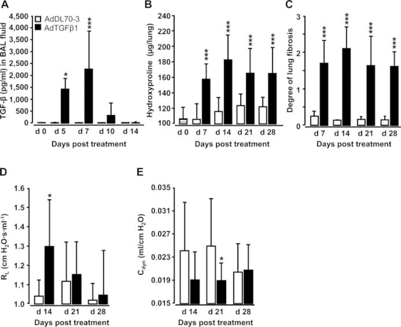
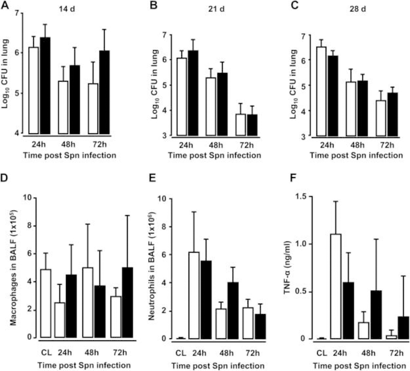
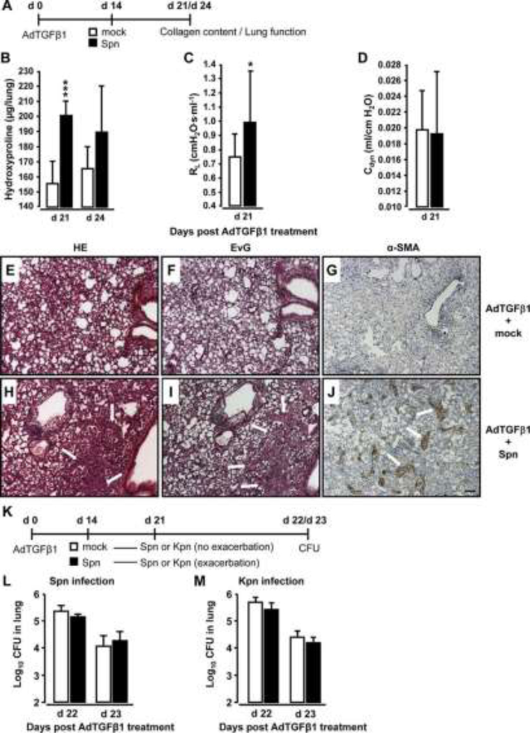
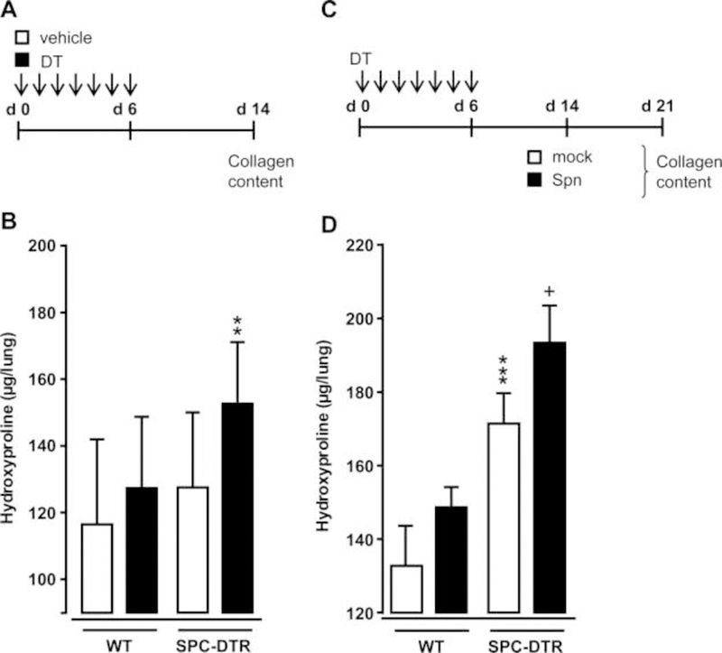
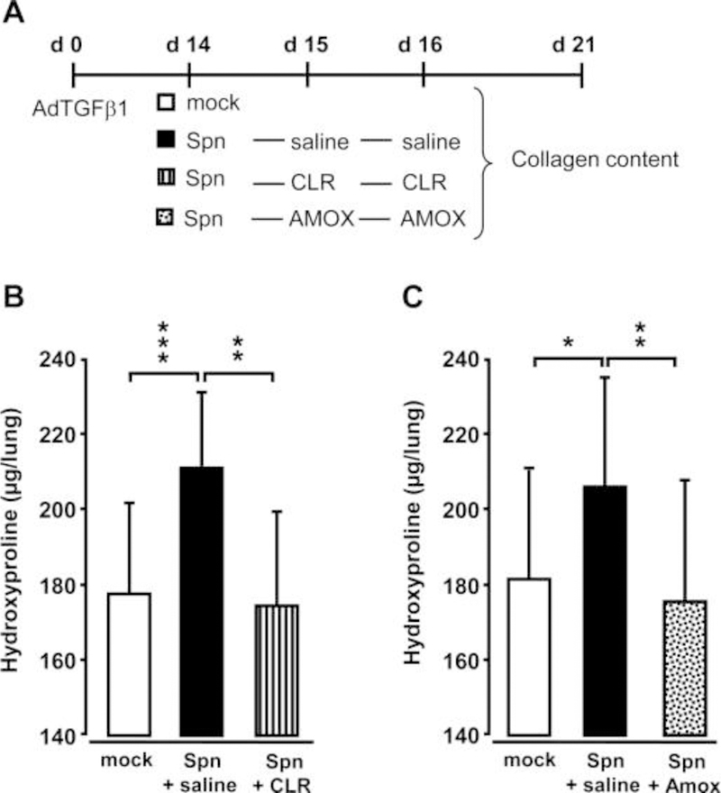
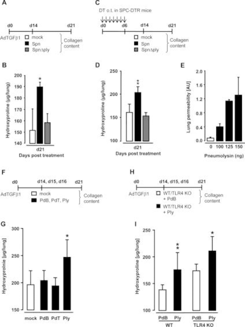
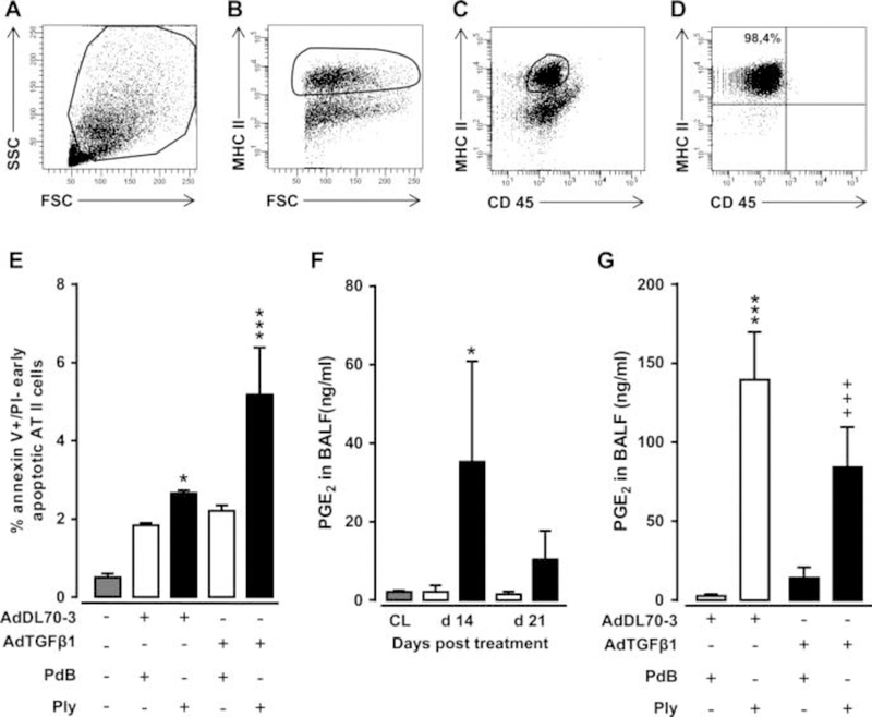
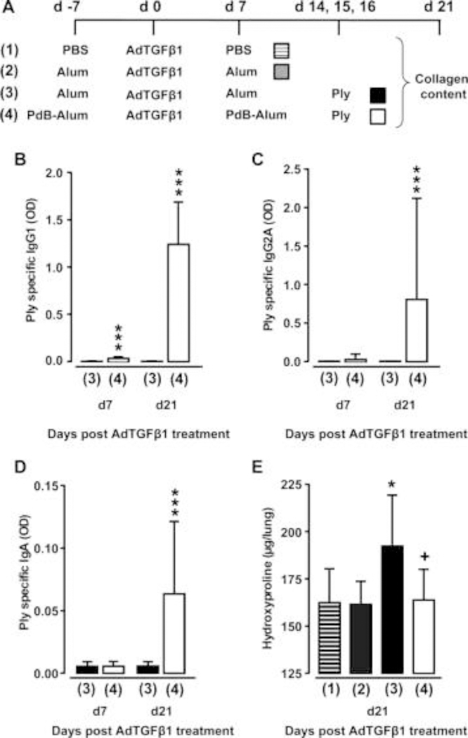
Measurements and main results: We demonstrate that wild-type mice exposed to adenoviral vector delivery of active transforming growth factor-β1 (TGFß1) or diphteria toxin (DT) treatment of transgenic mice expressing the DT receptor (DTR) under control of the surfactant protein C (SPC) promoter (SPC-DTR) to induce pulmonary fibrosis developed progressive fibrosis following infection with Spn, without exhibiting impaired lung protective immunity against Spn. Antibiotic treatment abolished infection-induced fibrosis progression. The cytotoxin pneumolysin (Ply) of Spn caused this phenomenon in a TLR4-independent manner, as Spn lacking Ply (SpnΔply) failed to trigger progressive fibrogenesis, whereas purified recombinant Ply did. Progressive fibrogenesis was also observed in AdTGFβ1-exposed Ply-challenged TLR4 KO mice. Increased apoptotic cell death of alveolar epithelial cells along with an attenuated intrapulmonary release of antifibrogenic prostaglandin E2 was found to underlie progressive fibrogenesis in Ply-challenged AdTGFβ1-exposed mice. Importantly, vaccination of mice with the non-cytotoxic Ply derivative B (PdB) substantially attenuated Ply-induced progression of lung fibrosis in AdTGFβ1-exposed mice.

Conclusions: Our data unravel a novel mechanism by which infection with Spn through Ply release induces progression of established lung fibrosis, which can be attenuated by protein-based vaccination of mice.

Keywords: Bacterial Infection; Idiopathic pulmonary fibrosis; Respiratory Infection.


Figures

Figure 1.
Figure 1.. Characterisation of AdTGFβ1-induced lung fibrosis in mice.
Naïve mice (lined bars, day 0 time points) were treated with control vector AdDL70-3 (1×10⁸ PFU, white bars) or AdTGFβ1 (1×10⁸ PFU, black bars) for various time points, as indicated. (A) Kinetics of active TGFβ1 protein in BAL fluid (BALF). (B) Analysis of hydroxyproline as a measure of lung collagen content in lung tissue. (C) Degree of lung fibrosis determined by histomorphometry/Ashcroft scoring. (D and E) Lung function in AdTGFβ1 and control-vector-exposed mice assessed by resistance (RL, D) and dynamic compliance (Cdyn, E). Data are shown as mean±SD of n=5 mice per time point and treatment group (n=10 mice per treatment group in D and E) and are representative of three (A–C) or two independently performed experiments (D and E). Data were analysed by Student’s t test. *p<0.05, ***p<0.001 compared with control-vector-treated mice. PFU, plaque-forming unit.
Figure 2.
Figure 2.. Effect of AdTGFβ1 on lung protective immunity against bacterial challenge.
(A–C) Bacterial loads were determined in lung tissue at 24, 48 and 72 h after Spn infection (1×10⁷ CFU/mouse) from mice exposed to AdTGFβ1 (black bars) or control vector (white bars) for 14 days (A), 21 days (B) or 28 days (C), as indicated. (D–F) BAL fluid (BALF) was collected from untreated control mice (striped bars) or at 24, 48 and 72 h after Spn infection from mice exposed to AdTGFβ1 or control vector for 14 days and numbers of macrophages (D) and neutrophils (E) as well as TNF-α protein levels (F) were determined. Data are shown as mean±SD of n=4 mice per time point and treatment group, and are representative of three independently performed experiments. Data were analysed by Mann–Whitney U test. CL, untreated control mice; CFU, colony-forming unit.
Figure 3.
Figure 3.. Bacterial infection triggers progressive pulmonary fibrosis in AdTGFβ1-exposed mice.
(A) Experimental profile. Mice were exposed to AdTGFβ1 (1×10⁸ PFU) for 14 days and were then either mockinfected (white bars) or Spn-infected (1×10⁷ CFU, black bars) for 7 or 10 days, as indicated. (B) Lung collagen content in Spn-infected mice at day 7 and 10 post-Spn challenge. (C and D) Assessment of resistance (RL, C) and dynamic compliance (Cdyn, D) in the lungs of AdTGFβ1-treated, Spn-infected mice on day 21 post- AdTGFβ1 (corresponding to day 7 post-Spn). (E–J) Histopathology of lungs from AdTGFβ1-treated mice subsequently challenged with mock (E–G) or Spn (H–J) for 7 days. Lung tissue sections were stained with H&E (E and H), Elastica-van-Gieson (EvG; F and I), and α-smooth muscle actin (α-SMA; G and J). Note substantially increased collagen deposition and myofibroblast accumulation in H, I and J (marked by white arrows). (K) Experimental profile to determine lung protective immunity of mice exhibiting Spn-induced progression of AdTGFβ1-induced lung fibrosis. On day 14 post-AdTGFβ1 (1×10⁸ PFU), mice were either mock-infected (white bars) or Spn-infected (1×10⁷ CFU, black bars) for 7 days, resulting in fibrosis progression. Subsequently, mice were reinfected with Spn (1×10⁷ CFU, L) or Kpn (5×10⁶ CFU, M) on day 7 after initial mock or primary bacterial infection, and bacterial loads were determined, as indicated (L and M). The data are shown as mean±SD of n=5 mice per time point and treatment group. The experiment was repeated twice with similar results. Data were analysed by Student’s t test (B–D) and Mann–Whitney U test (L and M). *p<0.05, ***p<0.001 compared with mock-treated mice. (E–J) Scale bar, 100 μm. CFU, colony-forming unit; PFU, plaque-forming unit.
Figure 4.
Figure 4.. Bacterial infection triggers progressive pulmonary fibrosis in diphtheria toxin (DT)- exposed transgenic SPC-DTR mice.
(A and B) Characterisation of DT-induced lung fibrosis in transgenic SPC-DTR mice. Transgenic SPC-DTR mice and their wild-type (WT) littermates were either treated o.t. with vehicle (PBS) or DT daily for 7 days. (B) Lung collagen content in WT and transgenic SPC-DTR mice at day 14 after DT treatment. (C) Experimental profile. SPC-DTR mice and their WT littermates received daily o.t. applications of DT for 7 days and were then challenged with mock versus Spn on day 14, that is, the time of established lung fibrosis. (D) Lung collagen content in WT and SPC-DTR mice treated with DT followed by mock infection or Spn infection, as indicated in (C). The data are shown as mean±SD of n=5 mice per time point and treatment group, and is representative of two independently performed experiments. Data were analysed by two-way ANOVA. **p<0.01, compared with vehicle-treated SPC-DTR mice (B), ***p<0.001 compared with DT-treated WT mice, +p<0.05 compared with mock-infected SPC-DTR mice (D). ANOVA, analysis of variance; o.t., orotrachea; PBS, phosphate-buffered saline.
Figure 5.
Figure 5.. Effect of antibiotic treatment on Spn-induced exacerbation of lung fibrosis in mice.
(A) Experimental profile. Mice were pretreated with AdTGFβ1 (1×10⁸ PFU/mouse) for 14 days and were then either mock-infected (white bars) or Spn-infected (1×10⁷ CFU/mouse). Subsequently, mice received saline (black bars), clarithromycin (CLR, 50 mg/kg b.w., striped bars) or amoxicillin (AMOX, 100 mg/kg b.w., dotted bars) on day 1 and 2 post-infection (corresponding to days 15 and 16 post-AdTGFβ1). (B and C) Lung collagen contents in the lungs of mice of the respective treatment groups determined on day 21 post-AdTGFβ1. The data are shown as mean±SD of n=3 mice per time point and treatment group, and is representative of four independently performed experiments. Data were analysed by one-way ANOVA. *p<0.05, **p<0.01, ***p<0.001 compared with Spn-infected + saline-treated mice. ANOVA, analysis of variance; b.w., body weight; CFU, colony-forming unit; PFU, plaque-forming unit.
Figure 6.
Figure 6.. Effect of pneumolysin on progression of AdTGFβ1 and SPC-DTR-dependent lung fibrosis.
(A) Experimental profile. Mice were exposed to AdTGFβ1 (1×10⁸ PFU/mouse) for 14 days and were then either mock-infected (white bars) or Spn-infected (1×10⁷ CFU/mouse, black bars) or were infected with the isogenic mutant strain of Streptococcus pneumoniae SpnΔply (1×10⁷ CFU/mouse, grey bars). (B) On day 21, lungs were harvested and lung hydroxyproline contents were determined. (C) Experimental profile. SPC-DTR transgenic mice received daily DT applications for 7 days orotracheally and were then mock-infected (white bars) or Spn-infected (1×10⁷ CFU/mouse, black bars) or were infected with the isogenic mutant strain of S. pneumoniae SpnΔply (1×10⁷ CFU/mouse, grey bars) on day 14. (D) On day 21, lungs were harvested and lung hydroxyproline contents were determined. (E) Lung permeability determined in naïve wild-type (WT) mice (white bar) or mice at 6 h after single challenge with Ply (black bars), as indicated. (F) Experimental profile for repetitive treatment of AdTGFβ1-exposed WT mice with Ply. Mice were exposed to AdTGFβ1 (1×10⁸ PFU/mouse) for 14 days, and on days 14, 15 and 16, mice received PBS o.t. (50 μL, white bars) or either PdB or PdT or Ply (∼50 haemolytic units per mouse) in 50 μL PBS (black bars), as indicated. (G) On day 21, lungs were harvested for determination of collagen contents. (H) Experimental profile. WT and TLR4 KO mice were exposed to AdTGFβ1 (108 PFU/mouse) for 14 days, and on days 14, 15 and 16, mice received PdB (white bars) or Ply (∼50 haemolytic units per mouse, black bars) o.t. for three consecutive days. (I) On day 21, lungs were harvested for determination of collagen contents. The data are shown as mean±SD of n=5 mice per time point and treatment group and are representative of two independently performed experiments. Data were analysed by one-way ANOVA (B, D, G) and two-way ANOVA (I). *p<0.05, **p<0.01 compared with mock and SpnΔply treatment in (B, D), and compared with mock or PdB or PdT treatment in (G), and compared with PdB treatment (I). ANOVA, analysis of variance; CFU, colony-forming unit; o.t., orotracheal; PBS, phosphatebuffered saline; PdB, pneumolysin derivative B; PdT, pneumolysin derivative T; PFU, plaque-forming unit.
Figure 7.
Figure 7.. Effect of repetitive Ply challenge of AdTGFβ1-exposed mice on apoptosis and PGE2 levels of AT II cells.
(A–D) Gating strategy for flow sorting of AT II cells isolated on day 21 from the lungs of mice exposed to AdTGFβ1 for 14 days followed by repetitive PdB or Ply challenge, as outlined in the legend to figure 6F. (A–D) Pre-enriched AT II cells were stained with anti-MHCII and anti-CD45 antibodies in the presence of anti-CD16/32 Ab, and were then gated according to their FSC-A versus SSC-A characteristics (A), followed by hierarchical subgating according to their FSC versus MHC II (B) and CD45 versus MHC II (C) expression. (D) Post-sort analysis of sorted AT II cells revealed sort purities of >98%, as verified by modified Papanicolaou stain and electron microscopic detection of lamellar bodies (data not shown). (E) Percent of early apoptotic (annexin Vpos, propidium iodideneg) AT II cells purified from the lungs of untreated mice (grey bars), or mice challenged with control vector (white bars) or AdTGFß1 (black bars) for 14 days and PdB or Ply for 7 days, as outlined in the legend to (E). (F) PGE2 levels in BAL fluids of untreated mice (grey bars) or mice treated with control vector (white bars) or AdTGFβ1 (black bars) for the indicated time intervals. (G) PGE2 levels in BAL fluids of mice treated with control vector or AdTGFβ1 for 14 days, followed by repetitive treatment with PdB versus Ply on days 14, 15 and 16 post-vector treatment, as indicated. PGE2 levels were determined on day 17 after vector treatment. The data are shown as mean±SD of n=3 mice per time point and treatment group (n=5 mice per time point and treatment group in F), and are representative of two independently performed experiments. Data were analysed by two-way ANOVA (E and G) or Student’s t test (F). *p<0.05, ***p<0.001 compared with control vector, +PdB treatment and +++p<0.001 compared with control vector +Ply treatment. ANOVA, analysis of variance; FSC, forward scatter; PdB, pneumolysin derivative B.
Figure 8.
Figure 8.. Effect of vaccination on Ply-induced fibrosis progression.
(A) Experimental profile. On day -7, mice received PBS or Alum or PdB-Alum (intraperitoneal, 10 μg/mouse). On day 0, mice were exposed to AdTGFβ1, followed by re-immunisation with PdB-Alum (20 μg/mouse) or i.p. injection of Alum only or PBS on day 7 post-vector treatment, as indicated. On days 14, 15 and 16 after AdTGFβ1 exposure, mice received either PdB or Ply o.t., as indicated. (B–D) Plasma antibody titres of Ply specific IgG1 (B; 1:3000 diluted) or IgG2A (C; 1:30 diluted) or IgA (D; 1:30 diluted) from groups of mice as outlined in (A). (E) Lung hydroxyproline levels in experimental groups of mice as outlined in (A). The data are shown as mean±SD of n=9 mice per time point and treatment group, and are representative of three independently performed experiments. Data were analysed by Mann–Whitney U test (B–D) or one-way ANOVA (E). *p<0.05 compared with group 2, ***p<0.001 compared with group 3, +p<0.05 compared with group 3. ANOVA, analysis of variance; o.t., orotrachea; PBS, phosphate-buffered saline; PdB, pneumolysin derivative B.

Similar articles

Cited by

References

    1. Raghu G, Collard HR, Egan JJ, et al. An official ATS/ERS/JRS/ALAT statement: idiopathic pulmonary fibrosis: evidence-based guidelines for diagnosis and management. Am J Respir Crit Care Med 2011;183:788–824. - PMC - PubMed
    1. King TE Jr., Pardo A, Selman M. Idiopathic pulmonary fibrosis. Lancet 2011;378:1949–61. - PubMed
    1. McMillan TR, Moore BB, Weinberg JB, et al. Exacerbation of established pulmonary fibrosis in a murine model by gammaherpesvirus. Am J Respir Crit Care Med 2008;177:771–80. - PMC - PubMed
    1. Martinez FJ, Safrin S, Weycker D Jr., et al. The clinical course of patients with idiopathic pulmonary fibrosis. Ann Intern Med 2005;142(12 Pt 1):963–7. - PubMed
    1. Collard HR, Moore BB, Flaherty KR Jr., et al. Acute exacerbations of idiopathic pulmonary fibrosis. Am J Respir Crit Care Med 2007;176:636–43. - PMC - PubMed

Publication types

MeSH terms