. 2015 May 26;112(21):6601-6.
doi: 10.1073/pnas.1424638112. Epub 2015 May 11.

N-lactoyl-amino acids are ubiquitous metabolites that originate from CNDP2-mediated reverse proteolysis of lactate and amino acids

Affiliations

N-lactoyl-amino acids are ubiquitous metabolites that originate from CNDP2-mediated reverse proteolysis of lactate and amino acids

Robert S Jansen et al. Proc Natl Acad Sci U S A. .

Abstract

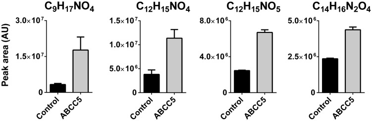
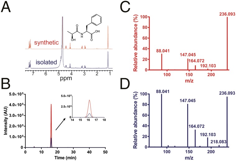
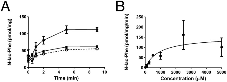
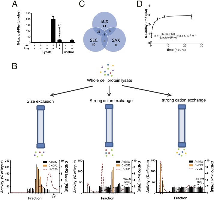
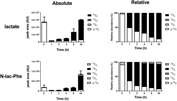
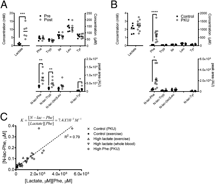
Despite technological advances in metabolomics, large parts of the human metabolome are still unexplored. In an untargeted metabolomics screen aiming to identify substrates of the orphan transporter ATP-binding cassette subfamily C member 5 (ABCC5), we identified a class of mammalian metabolites, N-lactoyl-amino acids. Using parallel protein fractionation in conjunction with shotgun proteomics on fractions containing N-lactoyl-Phe-forming activity, we unexpectedly found that a protease, cytosolic nonspecific dipeptidase 2 (CNDP2), catalyzes their formation. N-lactoyl-amino acids are ubiquitous pseudodipeptides of lactic acid and amino acids that are rapidly formed by reverse proteolysis, a process previously considered to be negligible in vivo. The plasma levels of these metabolites strongly correlate with plasma levels of lactate and amino acid, as shown by increased levels after physical exercise and in patients with phenylketonuria who suffer from elevated Phe levels. Our approach to identify unknown metabolites and their biosynthesis has general applicability in the further exploration of the human metabolome.

Keywords: ABCC5; MRP5; physical exercise; unknown metabolites; untargeted metabolomics.


Conflict of interest statement

The authors declare no conflict of interest.

Figures

Fig. 1.
Fig. 1.
Four unknown metabolites accumulate in culture medium of HEK 293 cells overexpressing ABCC5. HEK 293 control and HEK 293/ABCC5 cells were grown to confluence in six-well plates and cultured for an additional 3 d. Culture medium was analyzed using untargeted LC/MS metabolomics. Comparison of the metabolite profiles revealed four unknown metabolites that were significantly more abundant in medium of HEK 293/ABCC5 cells. Data are presented as mean plus SD (n = 3). AU, arbitrary units. The intracellular levels of the unknowns are depicted in Fig. S1.
Fig. 2.
Fig. 2.
Unknown C12H15NO4 is N-lac-Phe. (A) 1H-NMR spectrum of isolated C12H15NO4 recorded in D2O at 300 MHz matches the 1H-NMR spectrum of the synthesized reference N-lac-Phe. (B) Unknown C12H15NO4 elutes at the same time as N-lac-Phe. Extracted ion chromatogram of the mass corresponding to C12H15NO4 (m/z 236.09) in conditioned medium from HEK 293/ABCC5 cells before (depicted in blue) and after (depicted in red) spiking with synthesized N-lac-Phe (1 μg/mL, 4.2 μM). The high-resolution MS2 fragmentation spectrum of synthesized N-lac-Phe (C) and unknown C12H15NO4 in conditioned medium from HEK 293/ABCC5 (D) match. Differences in background and intensities can be attributed to the lower concentration of unknown C12H15NO4. Additional NMR and MS spectra are presented in Figs. S2 and S4, respectively.
Fig. 3.
Fig. 3.
N-lac-Phe is transported into inside-out membrane vesicles by ABCC5. (A) Control vesicles (▪) and ABCC5-containing vesicles (○ and ●) were incubated with 100 μM N-lac-Phe at 37 °C in the presence (solid line) and absence (dashed line) of 5 mM ATP. At the indicated time points, a sample containing 75 μg of protein was taken. After washing over a filter, the vesicular content was analyzed by LC/MS (n = 3–6). (B) Concentration dependence was assessed by incubating control and ABCC5-containing vesicles with several concentrations of N-lac-Phe in the presence of ATP and determining ABCC5-dependent uptake after 2 min (n = 3–4 for concentration <1,000 μM and n = 8 for concentration ≥1,000 μM). The data were fitted to Michaelis–Menten kinetics (solid line) using GraphPad Prism. Data are presented as mean ± SEM.
Fig. 4.
Fig. 4.
N-lac-Phe is formed by CNDP2. (A) N-lac-Phe is formed in the presence of Phe, lactate, and intact protein. Whole-cell lysate or control buffer [25 mM Tris⋅HCl (pH 7.4)] was incubated for 30 min at 37 °C in the presence or absence of 10 mM substrates. N-lac-Phe formation was determined by LC/MS and is expressed in arbitrary units. Data are presented as mean (n = 3) plus SD. (B) Whole-cell lysate was fractionated in parallel on three different columns. Enzyme activity was assessed by incubation with 10 mM lactate and Phe (30 min, 37 °C) and is normalized to the activity in unfractionated whole-cell lysate. The levels of CNDP2 were determined in active fractions and neighboring inactive control fractions (all marked by an asterisk) using LC/MS proteomics and are expressed as a peptide spectrum match (PSM) (n = 1). CV, column volume. (C) Although multiple proteins coeluted with enzyme activity for each single fractionation, only a single protein, CNDP2, coeluted with activity in all three fractionations. SAX, strong anion exchange; SCX, strong cation exchange; SEC, size exclusion chromatography. (D) Human recombinant CNDP2 (1 μg) was incubated (37 °C) with 10 mM lactate and Phe in 25 mM Tris⋅HCl (pH 7.4) containing 0.1 mM MnCl2. N-lac-Phe levels were determined by LC/MS and are expressed as arbitrary units. Data are presented as mean (n = 3) plus SD. Additional enzyme kinetics are presented in Figs. S5, S8, and S9.
Fig. 5.
Fig. 5.
13C-Metabolic labeling of lactate and N-lac-Phe occurs at a similar speed in HEK 293 cells. HEK 293 cells were grown to confluence in six-well plates, at which point the medium was replaced with medium containing 13C6-glucose. At several time points, the amount of lactate and N-lac-Phe containing only 12C (U-12C; unlabeled) or one to four 13C atoms per molecule (13C1–4) was determined in lysate by accurate mass LC/MS. Fully 13C-labeled lactate contains three 13C-atoms, which is reflected in the 13C3-labeling of N-lac-Phe. The minor presence of 13C1, 13C2, and 13C4 isotopologs can be explained by the natural occurrence of the 13C isotope (∼1% of all carbon atoms). Levels are expressed as absolute (arbitrary units) and relative values (percentage of isotope total). Data are presented as mean (n = 3) and SD (only for absolute values). Isotope data of corresponding culture medium samples are presented in Fig. S6.
Fig. 6.
Fig. 6.
N-lactoyl-amino acid levels in human plasma depend on lactate and amino acid concentrations. (A) Plasma was collected from human volunteers before (pre) and after (post) 5–10 min of strenuous exercise. Lactate and N-lactoyl-amino acid levels in human plasma rapidly increase after physical exercise, whereas the corresponding amino acid levels are unaffected. N-lactoyl-amino acid levels did not increase in resting controls. Horizontal bars represent means (n = 6). (B) Phe and N-lac-Phe levels are significantly increased in plasma of patients with PKU compared with controls, whereas plasma levels of other amino acids, N-lactoyl-amino acids, and lactate are similar. Horizontal bars represent means (n = 6 for controls, n = 11 for patients with PKU). (C) Plasma N-lac-Phe levels are in apparent equilibrium with plasma lactate and Phe levels. Plasma samples obtained from patients with PKU and controls, volunteers before and after exercise, and controls with high lactate due to prolonged whole-blood storage at ambient temperature were analyzed for lactate, Phe, and N-lac-Phe concentration as described in Materials and Methods (n = 34). The dotted line represents a fitted linear function, of which the slope is 7.4 × 10⁻²⋅M−1. N-lac-Phe is not formed in plasma (Fig. S7).

References

    1. Patti GJ, Yanes O, Siuzdak G. Innovation: Metabolomics: The apogee of the omics trilogy. Nat Rev Mol Cell Biol. 2012;13(4):263–269. - PMC - PubMed
    1. Tautenhahn R, et al. An accelerated workflow for untargeted metabolomics using the METLIN database. Nat Biotechnol. 2012;30(9):826–828. - PMC - PubMed
    1. Wishart DS, et al. HMDB 3.0—The Human Metabolome Database in 2013. Nucleic Acids Res. 2013;41(Database issue):D801–D807. - PMC - PubMed
    1. Fiehn O, Barupal DK, Kind T. Extending biochemical databases by metabolomic surveys. J Biol Chem. 2011;286(27):23637–23643. - PMC - PubMed
    1. Shin S-Y, et al. Multiple Tissue Human Expression Resource (MuTHER) Consortium An atlas of genetic influences on human blood metabolites. Nat Genet. 2014;46(6):543–550. - PMC - PubMed

Publication types